


STRUCTURAL ORGANISATION IN ANIMALS
INTRODUCTION
- Tissue is a group of similar cells along with intercellular substances that perform a specific function.
- Term tissue was used by Bichat (1972).
- All complex animals consist of only four basic types of tissues. These tissues are organised in specific proportion and pattern to form an organ like stomach, lung, heart and kidney etc.
- When two or more organs perform a common function by their physical and/or chemical interaction, they together form organ system, e.g., digestive system, respiratory system, etc.
- The tissues are different and are broadly classified into four types : epithelial, connective, muscular and neural.
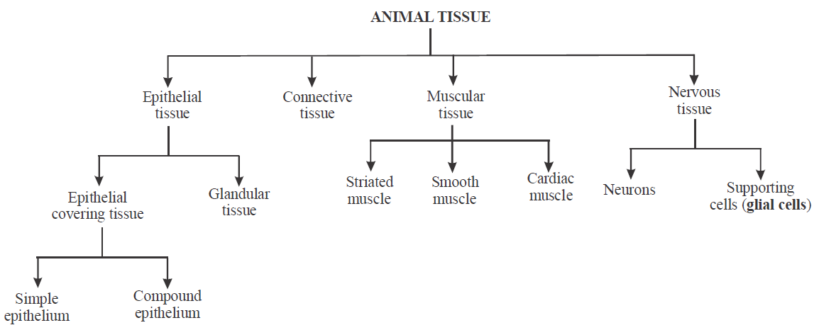
Flow chart : Animal Tissue
Table : Types of tissue on the basis of function and location.

EPITHELIAL TISSUE
- The word epithelium was introduced by Ruysch.
- The cells in epithelial tissue are very closely packed together and joined with little space between them.
- Common structures present in epithelial membrane are – intercellular junctions, basement membrane and structures on the free surface of cell like microvilli, cilia, flagella etc.
- All cells in epithelium are held together with little intercellular material.
- Specialised junctions provide both structural and functional links between its individual cells.
- Three types of cell junctions are - tight, adhering and gap junctions.
- Tight junctions help to stop substances from leaking across a tissue.
- Adhering junctions perform cementing to keep neighbouring cells together.
- Gap junctions facilitate the cells to communicate with each other by connecting the cytoplasm of adjoining cells, for rapid transfer of ions, small molecules and sometimes big molecules.
- The bottom layer cells rests on basement membrane.
- Basement membrane is composed of a network of fibres which includes collagen (in a matrix) and proteoglycans and sieves a selective filter determining which molecules diffuse from the undergoing connective tissue.
- Types of epithelial tissues are – simple epithelium and compound epithelium.
Table : Difference between simple epithelium and compound epithelium
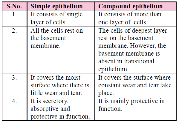
SIMPLE EPITHELIUM
- Simple epithelium is composed of a single layer of cells and every cell rests on the basement membrane.
- On the basis of structural modification, simple epithelium is divided into three types – squamous, cuboidal and columnar.
SQUAMOUS EPITHELIUM
- The squamous epithelium consists of a single thin layer of flattened cells with irregular boundaries.
- They occur in the walls of blood vessels and air sacs of the lungs.
- It looks like tiles of floor, hence called pavement epithelium.
CUBOIDAL EPITHELIUM
- The cuboidal epithelium consists of a single layer of cube-like cells.
- They are commonly found in ducts of glands and tubular parts of nephrons in the kidneys and its main functions are secretion and absorption.
- The epithelium of Proximal Convoluted Tubule (PCT) of nephron in the kidney has microvilli.
- Specialized cuboidal cells are capable of producing gametes as found in gonads. These are called germinal epithelium. E.g., ova (female gametes) and sperm (male gametes).
COLUMNAR EPITHELIUM
- The columnar epithelium consists of a single layer of tall and slender cells having basally located nuclei. Free surface may have microvilli.
- They are found in the lining of the stomach and intestine and help in secretion and absorption.
- Columnar epithelium is ciliated in bronchioles & fallopian tubes.
Their function is to move particles or mucus in a specific direction over the epithelium.
- Some of columnar or cuboidal cells get specialised for secretion and are called glandular epithelium. They are mainly of two types: unicellular, consisting of isolated glandular cells (goblet cells of the alimentary canal), and multicellular, consisting of a cluster of cells (salivary gland).
COMPOUND EPITHELIUM
- Compound epithelium is made of more than one layer (multi-layered) of cells.
- It has a limited role in secretion and absorption. They provide protection against chemical and mechanical stresses. It is also known as stratified epithelium.
- They cover the dry surface of the skin, the moist surface of buccal cavity, pharynx, inner lining of ducts of salivary glands and of pancreatic ducts.
- Different types of compound epithelium are
- stratified squamous keratinized epithelium
- stratified squamous non-keratinized epithelium
- stratified columnar epithelium
STRATIFIED SQUAMOUS KERATINIZED EPITHELIUM
- Stratified squamous epithelium is characterized by multiple layers of cells with typical flattened squamous cells at the free or outer surface of the sheet.
- The presence of keratin in these cells contributes to the protective qualities of skin covering the body surface.
- Keratin is dead and waterproof so it protects the underlying tissues from abrasion and infection. E.g., epidermis of the skin of land vertebrates.
STRATIFIED SQUAMOUS NON-KERATINIZED EPITHELIUM
- Its free surface is moist, and the outer epithelial cells, unlike those found in the skin, do not contain keratin.
- This type of epithelium serves a protective function. It is found lining the oral cavity (buccal cavity), pharynx, oesophagus, anal canal, lowerpart of urethra, vocal cords, vagina, cervix (lower part of uterus) and cornea of eyes.
STRATIFIED COLUMNAR EPITHELIUM
- It is a protective epithelium and has multiple layers of columnar cells, only the most superficial cells are truly columnar in appearance.
- Epithelium of this type is rare.
- It is found in male urethra and in the mucous layer near the anus. It also lines mammary gland, ducts and epiglottis.
NOTES
- Stratified transitional epithelium (Urothelium) : It contains cuboidal or columnar shaped cells, which are thin and stretchable.
- It lacks germinative layer, basement membrane. Stratified transitional epithelium is typically found in the body areas such as the wall of urinary bladder, ureter and renal pelvis. It is located in all the hollow viscera subjected to stress and protects organ wall from tearing.
- Neurosensory epithelium : Olfactory mucosa, called Schneiderian membrane, lining of internal nares, retina of eyes and epithelial covering of tongue containing taste buds are examples of neurosensory epithelia. The sensory cells bear, at their free ends, slender "sensory hairs" to receive specific stimuli. Basely, these cells are connected, by means of synapses, with fine fibrils of sensory nerves.
- Pigmented epithelium : The epithelial cells of the basal layer of retina contain pigment. Hence, this layer is often referred to as a pigmented epithelium. E.g., - Pigmented layer of retina, iris and skin.
CONNECTIVE TISSUE
- Connective tissues is the most abundant supporting tissue of the body.
- They are named connective tissues because of their special function of linking and supporting other tissues/organs of the body.
- About 30% of the body mass is formed of connective tissue.
- Connective tissue is divided into three categories - connective tissue proper (including matrix, cells & protein fibres), skeletal tissue (containing bones & cartilage), and vascular tissue (including blood & lymph).
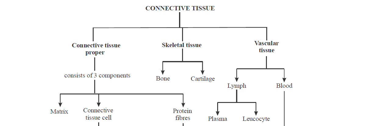
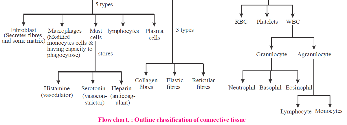
CONNECTIVE TISSUE PROPER
- Matrix accumulate between cells and fibres. Matrix is a non living material which may be liquid (e.g., blood), semisolid (e.g., connective tissue) and solid (e.g., bone).
- Matrix consists of mainly water and sulfated mucopolysaccharide.
- The cells of connective tissue are living and responsible for secreting the large amount of intercellular ground substance (matrix).
- Types of connective tissue cells are - fibroblast, macrophages, mast cells, lymphocytes and plasma cells.
- Connective tissue is composed of several protein fibres such as collagen, elastic and reticular fibres.
- Collagenous fibres are the most abundant fibrous element of areolar and other connective tissues. These are long, unbranched fibres of a soluble and shining collagen protein (tropocollagen). These fibres are more strengthful and provide maximum tensile strength. These are colourless and hyaline, yet called white fibres to distinguish them from yellow elastin fibres.
- Collagen protein is the most abundant protein of the body. It constitutes 25% of the total body protein.
- Collagen fibre can be stained by eosin. When collagen fibres are removed from the areolar tissue they become loose and elastic. E.g., Bone, cartilage, ligament and tendon.
- Elastic fibres are formed of elastin protein. These fibres are less numerous, thinner, branched, anastomosing, and of a pale yellow colour. These are very elastic and remain stretched due to tension in the areolar tissue, when broken in teased preparations, these coil and curl like tense wires.
- Elastin is probably the most resistant of all body proteins to chemical changes. Thousands of years old 'mummies' still have their arteries intact due to well-preserved elastin fibres. They are the orceinophilic i.e., stained by orcein.
- Reticular fibres are delicate, freely branching and inelastic fibres of reticulin protein, found interwoven, to form networks. These are very abundant in embryos, new born babies and in healing and regenerating wounds. In areolar tissues of adults, these are mostly replaced by collagen fibres, but remain abundant in lymphoid and blood forming tissues and in the stroma of pancreas, liver etc. They are stained with AgBr and AgNO3, hence are called argentophilic or argyrophilic.
- On boiling collagen and reticular fibres, both convert in glue.
TYPES OF CONNECTIVE TISSUE PROPER
On the basis of cells & fibres present, connective tissue is further divided into - areolar connective tissue, adipose connective tissue, dense connective tissue (white fibrous connective tissue & yellow elastic connective tissue), reticular connective tissue, mucoid connective tissue and pigmented connective tissue.
I. Areolar connective tissue
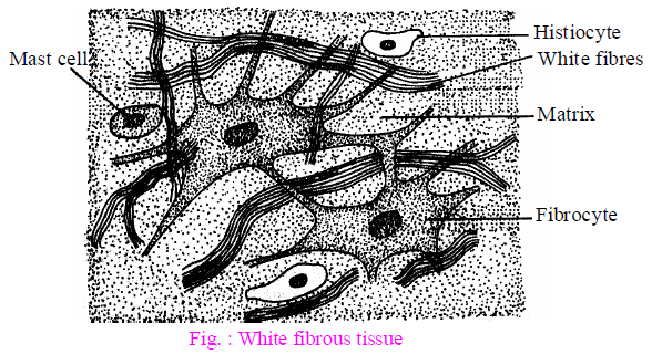
- Loose or areolar connective tissue has cells and fibres loosely arranged in a semi-fluid ground substance. For example, areolar tissue is present beneath the skin. It serves as a support framework for epithelium. It contains fibroblasts (cells that produce and secrete fibres), macrophages and mast cells.
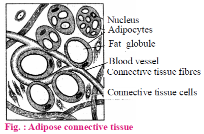
II. Adipose connective tissue
- Adipose tissue is a modified form of areolar tissue, made up of specialized large spherical fat cells (below the skin) or adipocytes. Adipose tissue is located mainly beneath the skin, in bone marrow, kidney, liver etc. The excess of nutrients which are not used immediately are converted into fats and are stored in this tissue.
- Adipose tissue chifley act as "food reserves" or fat depots for storage and metabolism of lipids. Besides this, they also act as heat insulators and pressure, pull and push absorbers.
III. Dense connective tissue
- Fibres and fibroblasts are compactly packed in the dense connective tissues. Orientation of fibres show a regular or irregular pattern and are called dense regular and dense irregular tissues.
- In the dense regular connective tissues, the collagen fibres are present in rows between many parallel bundles of fibres. Dense regular connective tissue are found in flexible ligament.
- Dense irregular connective tissue has fibroblasts and many fibres (mostly collagen) that are oriented differently. This tissue is present in the skin.
- Dense connective tissue are of two types – yellow elastic & white fibrous.
- Yellow elastic fibrous tissue : The matrix is with numerous and closely packed yellow or elastin fibres which are similar to but thicker than those of areolar connective tissue. It is elastic and flexible. It forms wall of blood vessels, lungs, true vocal cords, trachea, capsule of spleen and bronchioles. It also forms sheet in ligaments. Ligaments is a modified yellow elastic fibrous tissue and connects bone to bone.
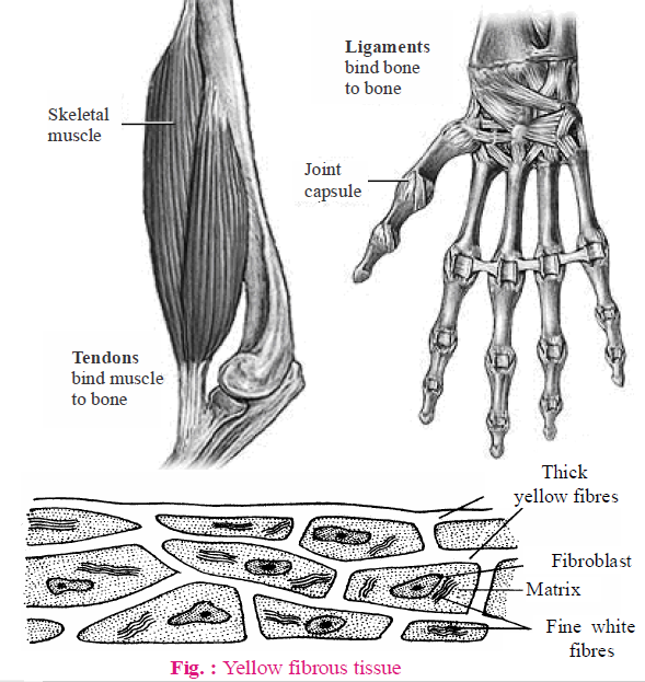
- White fibrous tissue is modified form of areolar tissue. Only collagen fibres are present in the matrix and cells are mainly fibroblasts. It is present at the joints between skull bones and makes them immovable. It is also found in the dermis of higher mammals.
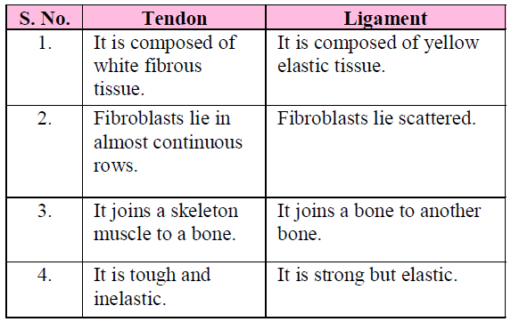
- Tendon (which connects skeletal muscle to a bone) is a modification of white fibrous tissue.
Table : Difference between Tendon and Ligament
IV. Reticular connective tissue
Reticular connective tissue is a network of reticular fibres that form a soft skeleton to support the lymphoid organs (lymph nodes, bone marrow and spleen).
V. Mucous connective tissue
Mucous connective tissue is composed of gelatinous substance (called wharton's jelly), few fibroblast and collagen. It is found in the umbilical cord and vitreous humor.
VI. Pigmented connective tissue
Pigmented connective tissue gives colours to the structures. They are found in the iris of the eye and dermis of the human skin.
SKELETAL CONNECTIVE TISSUE
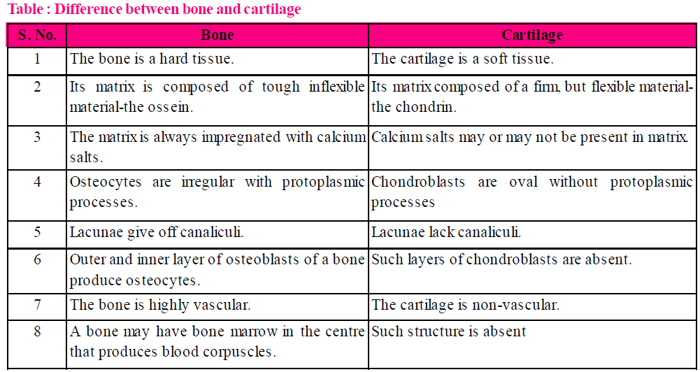
CARTILAGE
- The intercellular material of cartilage is solid and pliable and resists compression.
- Cells of this tissue (chondrocytes) are enclosed in small cavities within the matrix secreted by them. Most of the cartilage in vertebrate embryos are replaced by bones in adults.
- Cartilage is found in the tip of nose, outer ear joints, between adjacent bones of the vertebral column, limbs and hands in adults.
- Cartilage is avascular and nutrients are diffused through the matrix.
- The matrix of cartilage consists of glycoprotein material chondroitin sulfate and keratin sulfate.
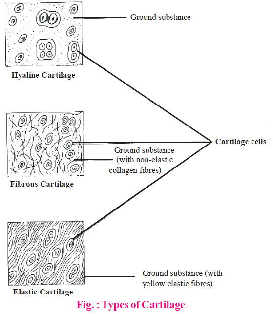
- There are three main types of cartilage - hyaline, elastic and fibrocartilage.
- Hyaline cartilage is an elastic, compressible tissue, located at the end of bones, on nose, bronchi, larynx etc.
- Inside the bone, hyaline cartilage serving as a centre of ossification or bone growth.
- Calcified cartilage is formed by the calcification of hyaline cartilage. It is found in the suprascapular & pubis in frog.
- Elastic cartilage helps to maintain the shape and flexibility of the organ and also strengthens and supports these structures.
- Yellow elastic cartilage is found in external ear and epiglottis.
- Fibrous cartilage is the strongest cartilage due to the presence of collagen fibres. It occurs in pubic symphysis of man and intervertebral disc. Fibrocartilage lacks perichondrium.
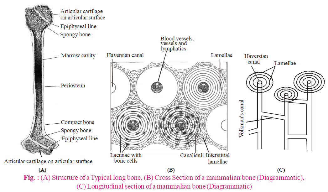
BONES
- Bone is a type of hard endoskeleton connective tissue.
- It is rich in calcium salts and collagen fibres which give bone its strength.
- Bone is the main tissue that provides structural frame to the body. Bones support and protect soft tissues and organs.
- The bone cells (osteocytes) are present in the spaces called lacunae.
- Bone cells are of three types – Osteoblasts (bone forming cells), osteocytes (bone maintaining cells) and osteoclasts (bone cleaning cells).
- Osteoblasts secretes collagen fibres and matrix of bone and are responsible for calcification of the matrix.
- Osteocytes are inactive bone cells. They are responsible for maintaining the matrix and can both secrete & resorb matrix.
- Osteoclasts destroy bone matrix and it is formed by the fusion of monocytes. They release lysosome, organic acids and hydrolytic enzymes to break down bone matrix.
- Periosteum is a membrane that forms an envelope around the bone.
Periosteum is comprised of two distinct layers– outer layer consists of thin white fibrous connective tissue and inner layer consist of osteoblasts.
- Bone matrix is composed of protein called ossein. The matrix forms thin plates called lamellae.
- Lamellae are of three types – Haversian lamellae (occur around Haversian canal), concentric or circumferential lamellae (inner to periosteum and outer to endosteum) and interstitial lamellae (between Haversian system). In the lamellae, minute bone cells osteocytes are present.
- Bones are of two types – compact bone & spongy bone.
- Compact bones form shaft of long bones & consists of yellow bone marrow.
- Spongy bones (also known as cancellous bone) carries no haversian system and consists of thin interconnecting bony struts called trabeculae. Spongy bones occurs in embryo, growing organisms & the swollen ends of long bones.
- Bone marrow is a specialized type of soft, diffuse connective tissue called "Myeloid tissue". It takes part in production of blood cells hence known as haemopoietic tissue.
- Endosteum is a membrane which lines the marrow cavity.
Bone marrow is composed of adipose tissue, areolar tissue and blood.
It is of two types – red bone marrow and yellow bone marrow.
(1) Red bone marrow
- It is red in colour due to the presence of a lot of blood vessels.
- In foetal life and at birth, it is present in the entire skeleton. After 5th year red bone marrow is replaced by yellow bone marrow, at 20-25 years red bone marrow is present at ribs, sternum, clavicles, vertebrae, scapula, pelvis, epiphysis of humerus and femur.
- It produces RBCs, WBC, platelets, granular leukocytes like basophils, eosinophils and neutrophils.
(2) Yellow bone marrow
- It is yellow in colour and has much fatty tissue (adipose tissue).
- It is present in the shaft of long bones.
- It produces blood cells in emergency i.e., at the time of excessive loss of blood, yellow bone marrow may be replaced by red bone marrow in anaemia.
VASCULAR TISSUE
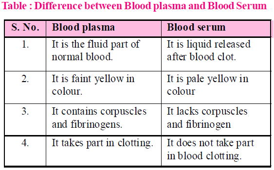
BLOOD
- Blood is a fluid connective tissue containing plasma, red blood cells (RBC), white blood cells (WBC) and platelets. It is the main circulating fluid that helps in the transport of various substances.
- Plasma constitutes about 5% of body weight. It represents matrix of blood. Plasma is slightly alkaline and transparent. It forms 55-60% by volume of blood.
- Blood corpuscles form 40-50% of the blood and are of three types viz. red blood corpuscles, white blood corpuscles and platelets.
Red blood corpuscles (RBCs or Erythrocytes)
- These occur only in vertebrates and are the most abundant (99%) of blood corpuscles, imparting the characteristic red colour to the blood.
- The shape, size and structure of RBCs vary in different types of vertebrates, but their function is the same in all, i.e. transport respiratory gases, especially the oxygen (O2).
- Each RBC is bounded by a dynamic, enzyme-containing plasma membrane. In human RBC, about 26.5 crore molecules of haemoglobin are packed in the intracellular framework. Water constitutes about 60% of RBC. The rest is solid. Haemoglobin forms about 34% of wet and 90% of dry weight of an RBC. Thus, 100 ml of normal human blood contains about 15 gm of haemoglobin on an average. An apparatus named haemoglobinometer is used to determine the haemoglobin content of blood.
- Function of RBCs : The major function of erythrocytes is to receive O2 of respiratory surfaces and then transport and readily deliver it to all cells of the body. This important function is performed by haemoglobin which has a great ability to combine loosely and reversibly with O2 and is, hence, called "respiratory pigment".
- Haemoglobin, in annelids, is dissolved in the plasma because of absence of red blood corpuscles. In mollusc and some arthropods, etc., a different respiratory pigment, haemocyanin is found dissolved in the plasma. This pigment is bluish due to the presence of copper in place of iron.
- ESR : It is called erythrocyte sedimentation rate. This test is measured by "Wintrobe's tube" and "Western blotting" method. It is the rate of sinking/settling down of RBC in the plasma to form rouleaux. Man has lower ESR as compared to women and it is lowest in newborn. Normal value of ESR in male is about 5 mm and in female 10 mm in first hour. A rise in ESR indicates the presence of infective/ destructive/ inflammatory diseases.
- Average life of RBC is 120 days.
- RBCs of frog : Amphibian RBCs are largest amongst the vertebrates. Those of Amphiuma and Proteus are largest amongst amphibians (about 82 µm). These are flattened and oval, disclike, but slightly biconvex due to a large oval and centrally-placed nucleus.
- RBCs of mammals : Mammals have smallest RBCs amongst the vertebrates. Those of musk deer are the smallest amongst the mammals, whereas the RBCs of other vertebrates are oval and nucleated, those of mammals are roughly circular (except those of the family camellidae - camels, llamas, dromedaries - which are oval in shape) and non-nucleated.
- RBCs of human : They are about 7.4µm in diameter and its thickness is 1 to 1.5µm. It is pale yellow in colour but appear to be red in group. Surface area of all RBCs of a person totals about 1500 to 2000 times the surface area of the body itself.
White blood corpuscles (WBCs or Leucocytes)
- They are nucleated, colourless and complete cells.
- They are bigger than RBC but their number is less. The number of WBC is 5,000 to 10,000 per cubic mm.
- WBC shows least constancy in shape.
- They are formed in red bone marrow, spleen, thymus and lymph nodes from myelocytes.
- The life of WBC is of 15 hours to 2 days. The WBC are destroyed outside the blood vessels and the process by which it comes out is called as diapedesis.
- An increase in the number of white blood corpuscles is called leukocytosis. More than 20,000 per cubic mm. indicates some disease. A decrease below 5000/Cu.mm is called leukopenia as in typhoid fever.
- WBC help to defend the body against infectious disease and foreign materials as part of the immune system.
Blood platelets
These are protoplasmic disc that are found in mammalian blood (lower vertebrates have spindle-shaped cells named thrombocytes). Platelets arise as detached tips of protoplasmic processes extending from the cytoplasm of giant cells, megakaryocytes of red bone marrow. The shape is oval to round, often stellate. There are approximately 300,000 platelets in a cubic millimetre of blood. Platelets are non-nucleated. Life span is about 5-9 days. Blood platelets helps in coagulation of blood by producing platelet factors (such as thromboplastin).
LYMPH
- Lymph can be defined as blood minus RBCs but more WBCs. Lymph is chiefly made of plasma plus leukocytes.
Most important centre for the formation of lymph is interstitial space.
- Interstitial fluid, intercellular fluid, tissue fluid and lymph all are same in composition.
Exchange of materials between blood and tissue fluid occurs through blood capillaries.
- Functions of lymph : The basic function of lymph is to bring back, into the vascular circulation, the cell debris, large colloid particles and the part of the blood plasma that had diffused out from arterial capillaries into the tissue fluid but has failed to return back into venous capillaries. The white corpuscles of the lymph are the same as those of the blood and have the same functions of defense and of assistance in tissue repair and healing. In intestinal wall, lymph capillaries called lacteals, are specially meant for absorption of fats.
MUSCLE TISSUE
- Muscle tissue is made up of long, excitable cells or muscle fibres that are capable of considerable contraction.
- Muscular tissue carries out mechanical work by contracting which involves a shortening & thickening of its fibre.
- Two proteins-actin & myosin are part of the machinery & ATP is the immediate energy source for the contraction.
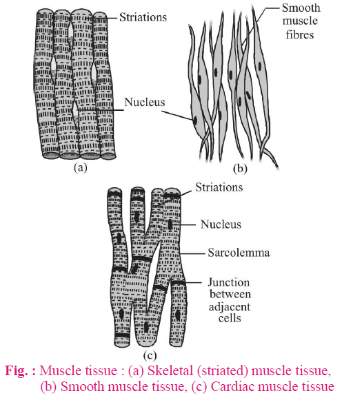
- On the basis of structure, location & function muscles are of three types – skeletal, smooth & cardiac muscles.
SKELETAL MUSCLE TISSUE
- Skeletal muscle tissue is closely attached to skeletal bones and controls motor movements & posture.
- Skeletal muscle work according to our will, hence are voluntary in nature.
- Skeletal muscle are known as striated muscles because their cells have transverse stripes.
- They are found in arms, hands, legs, body wall, tongue & upper part of pharynx.
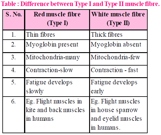
- Two types of fibres for skeletal muscles are - type I (slow twitch, red) and type II (fast twitch, white)
- These muscles are stimulated by acetylcholine.
SMOOTH MUSCLE TISSUE
- Cells of smooth muscle tissue are spindle shaped & pointed at their ends.
- Striation are absent due to different arrangement of actin & myosin filament.
- Cell junctions hold them together and they are bundled together in a connective tissue sheath.
- The wall of internal organs such as the blood vessels, stomach and intestine contains this type of muscle tissue.
- Smooth muscles are involuntary muscles as their functioning cannot be directly controlled.
- Smooth muscles may be multi unit (e.g., ciliary muscle, walls of large arteries) and single unit (e.g., alimentary canal, urinary bladder, uterus, oviducts etc.)
CARDIAC MUSCLE TISSUE
- Cardiac muscles are involuntary, cross striated & non-fatigued fibres.
- Fibres of these muscles contain large number of mitochondria & glycogen granules as they require large amounts of energy.
- They are richly supplied with blood.
- Cardiac muscle tissue is a contractile tissue present only in the heart forms the myocardium.
- Cardiac muscle never get fatigued because it rests & work for equal duration.
- Cell junctions fuse the plasma membranes of cardiac muscle cells and make them stick together. Communication junctions (intercalated discs) at some fusion points allow the cells to contract as a unit, i.e., when one cell receives a signal to contract, its neighbours are also stimulated to contract.
- Cardiac muscle tissue has no regenerative capacity.
NERVOUS TISSUE
- Nervous tissue is found in brain, spinal cord and nerves.
- It is responsible for coordinating and controlling many body activities.
- Nervous tissue is specialised to react to stimuli & to conduct impulses to various organs in the body which bring about a response to a stimulus.
- Cells of the nervous tissues are nerve cells (neurons) and supporting cells.
- Neurons forms the structural and functional unit of nervous tissue.
- Neurons are the largest cell in the body.
- Neurons consists of large cell body (cyton or soma), thin protoplasmic process - dendrite & a long process-axon.
- Supporting cells (also called glial cells) bind neurons together and insulate the neurons.
- Supporting cells, present in CNS, is divided into neuroglia, ependymal cells and neurosecretory cells.
ORGAN HISTOLOGY
LIVER
- It is the largest gland of the body and weighs 1-2 kg.
- It has maximum regeneration power (Compensatory regeneration).
- It is endodermal in origin and lies on the right side of the abdominal cavity.
- In humans, the liver is 4-lobed (Left Lobe, Right, Lobe, Quadrate Lobe and Caudate Lobe). In Frog and Rabbit it is 3 and 5 lobed respectively. The gallbladder is attached to the right lobe of the liver.
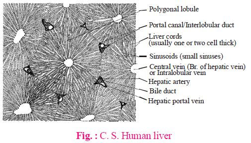
- The structural and functional unit of liver is lobule.
- Each lobule is surrounded by a connective tissue covering, called Glisson’s capsule (In human, the Glisson’s capsule is not developed). In the center of lobule there is a branch of hepatic vein, also known as intra-lobular vein. In between adjacent lobules, there are Interlobular ducts or portal canals containing branches of hepatic artery, lymphatic vessel, bile duct and hepatic portal vein. The hepatic cells (hepatocytes) are arranged in sheets, each 2-cells thick. Amongst the sheets of hepatic cells, there are blood lacunae and sinuses (sinusoids). The phagocytic cells, Kupffer Cells, lie in Glisson’s capsule and at the border of sinuses.
FUNCTIONS
- Phagocytosis - Kupffer cells are phagocytic cells and remove pathogens and toxic material from the blood.
- Excretion - Liver is not excretory organ but synthesis of urea, uric acid, creatinine etc. occurs in liver. The bilirubin (excretory product) is also formed in liver.
- Storage - It stores vitamin A, D, B12 , Iron (as ferritin), blood and glycogen etc.
- Haemopoesis - In foetus, the liver is a haemopoietic organ and produces RBCs, WBCs and platelets.
- Production - It produces bile juice, which is later on concentrated in gallbladder, and heparin, an anticoagulant. It also produces many plasma proteins like albumin, prothrombin and angiotensinogen. About 50% of the body lymph is produced in the liver. It synthesizes fatty acids, cholesterol and phospholipids also.
- It is also the seat of various metabolic reactions like Glycogenesis, Glycogenolysis, Gluconeogenesis, Deamination, Cori-cycle etc. It is also an important site for β-oxidation of fatty acids and degradation of cholesterol to form cholic acid of bile. It is the busiest organ of the body. It receives both oxygenated and deoxygenated blood. The blood supply to the liver in human is ~1500 ml per minute (1000 ml. from hepatic portal vein and 500 ml. from hepatic artery).
SPLEEN
- It is mesodermal in origin.
- It lies close to fundic stomach, in the left hand side of the abdominal cavity. (In frog, it is close to large intestine).
- Spleen is the largest lymphatic organ (15 cm. in diameter and 150 g. in weight)
- Unlike other lymphatic organs, it is not differentiated into cortex and medulla.
- The lymphatic sinuses and lymphatic vessels are also lacking in this organ.
- It consists of outer red-pulp (containing RBCs) and inner white-pulp (lacking RBCs). The nodules (germinal centres) in the white pulp synthesise lymphocytes.
FUNCTIONS
- Biological filter - It removes pathogens, like bacteria, and foreign material from the blood. Its phagocytic cells belong to reticulo-endothelial system.
- Blood Bank - It is called blood bank and stores blood in frog. In human, the maximum blood is, however, stored in liver.
- Haemopoietic organ - In human foetus and adult frog, the blood elements are manufactured in spleen. It also stores RBCs.
- Graveyard of RBCs - The worn out RBCs, whose lifespan is completed, are dumped in this organ.
- Immunological functions - It is the centre for lymphocyte production, which produce plasma cells and hence antibodies. It plays an important role in immunity.
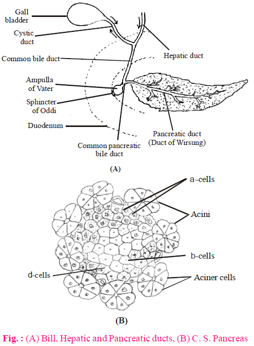
PANCREAS
- It, like liver, is endodermal in origin.
- It is the IInd largest digestive gland, only next to the liver, in the body (~ 15 cm in size)
- It lies in the coil of duodenum in the abdominal cavity.
- It is a Heterocrine (mixed) gland, containing both exocrine and endocrine parts.
- On the basis of structure, it is a compound tubulo-alveolar gland.
- Its exocrine part consists of a large number of Acini (sing. Acinus) which produce pancreatic juice.
- Pancreatic duct, before opening into the duodenum, joins Common bile duct and forms Ampulla of Vater, having ‘sphincter of Oddi’ at the common opening.
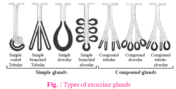
TYPES OF EXOCRINE GLANDS
On the basis of structure the exocrine glands can be differentiated into two broad categories, i.e. simple glands and compound glands :
(1) Simple glands
In such glands the duct (non-secretory part) is unbranched.
- Simple Unbranched glands - Their secretory part is also unbranched.
- Tubular - Uncoiled -e.g., Crypts of Lieberkuhn.
Coiled - e.g., Sweat glands. - Alveolar - e.g., Mucous and poison glands in Frog’s skin.
- Simple Branched glands - Their secretory part is branched.
- Tubular - e.g., Gastric glands.
- Alveolar - e.g., Sebaceous glands and Meibomian or Tarsal glands.
(2) Compound glands
In such glands, the duct (non-secretory part) is branched.
- Tubular - e.g., Non lactating mammary glands, Brunner’s glands.
- Tubulo-alveolar - e.g., Lactating mammary glands, parotid (salivary gland) and Pancreas etc.
- Alveolar - Submaxillary salivary glands
GUT (ALIMENTARY CANAL)
There are following 4-coatings (from inner to outer) in the gut lining.
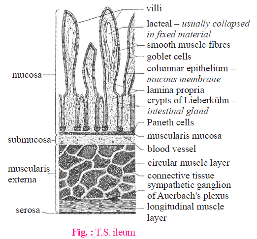
- Tunica mucosa - It is differentiated into epithelium, lamina propria and muscularis mucosa. It contains digestive glands. The epithelium in most part of the gut is simple columnar type. The lamina propria contains lymphatic tissue (GALT - Gut Associated Lymphatic Tissues) and various types of cells, like lymphocytes, plasma cells, eosinophils etc. It, however, lacks blood vessels and nerves.
- Tunica submucosa - It consists of connective tissue with blood vessels, nerve fibres and nerve plexuses (e.g., Meissner’s plexus).
- Tunica muscularis - It usually contains an inner layer of circular muscles and outer layer of longitudinal muscles. The Auerbach’s plexuses are usually present between these two layers of smooth muscles.
- Tunica externa - It consists of a thin layer of connective tissue covered with Visceral epithelium or Serosa (formed from coelomic mesoderm or peritoneum) and is attached to mesenteries.
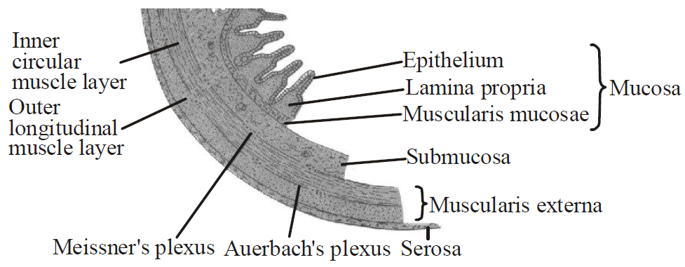
Fig. : General Plan of alimentary canal
OESOPHAGUS
- The epithelium or mucous membrane of tunica mucosa is thin and formed by stratified squamous cells.
- Tunica muscularis has voluntary (striated) muscles (upper 1/3rd part), voluntary and involuntary muscles (middle 1/3rd part) and involuntary (smooth) muscles (lower 1/3rd part).
STOMACH
- When stomach is empty, the tunica mucosa forms longitudinal folds called rugae.
- The gastric glands in tunica mucosa are simple tubular and branched. The neck of the glands contain mucus secreting cells; the body of the glands has chief (zymogen) and parietal (oxyntic) cells. The cells at the bottom are Argentaffin (enterochromaffin). The latter are endocrine cells of the stomach.
- The chief cells produce digestive enzymes.
- The parietal cells secrete intrinsic factor of Castle and HCl.
- Tunica muscularis of stomach contains inner oblique, middle circular (thickest) and outer longitudinal layer of smooth muscles.
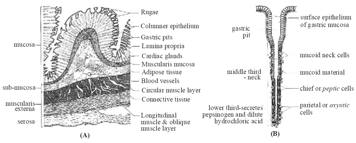
Fig. : (A) T. S. Stomach, (B) Fundic (Gastric) gland
SMALL INTESTINE
- This part is characterized by the presence of villi of various shapes.
- In small intestine, tunica mucosa consists of 4-types of cells.
- Enterocytes or absorptive cells - They have microvilli to form brush bordered epithelium and increase the surface area for absorption. They also secrete digestive enzyme.
- Goblet Cells - These are single celled, egg shaped glands and secrete mucus.
- Paneth Cells - They secrete digestive enzymes, for digesting the wall of bacteria.
- Argentaffin Cells - They are endocrine cells.
LARGE INTESTINE
- The villi are absent in this part of the intestine.
- In colon, the longitudinal muscles of tunica muscularis form 3-longitudinal bands, called teniae coli, to make colon sacculated.
- In anal canal, the mucous membrane is thrown into longitudinal folds, called Rectal columns of Morgagni.
INTEGUMENT
It is the part of the body wall and consists of skin and its derivatives (glands and exoskeleton)

The skin is ecto mesodermal in origin and consists of epidermis (ectodermal) and dermis (mesodermal).
MAMMALIAN SKIN
EPIDERMIS
It is stratified squamous epithelium and, from inner to our, consists of the following layers
- Stratum germinativum or stratum Malpighii (Innermost)
- Stratum spinosum
- Stratum granulosum
- Stratum lucidum
- Stratum corneum (outer/top most)
STRATUM GERMINATIVUM (STRATUM MALPIGHII)
- It is simple columnar epithelium.
- It is the only meristematic layer in epidermis (the mitotic divisions are faster during night).
- All other layers, cutaneous glands, and the exoskeleton develop from this layer.
- The pigment bearing cells, melanocytes, are present in between these cells.
STRATUM SPINOSUM
- It is a prickly layer with polyhedral cells bearing minute spine like structures.
STRATUM GRANULOSUM
- Its polygonal cells contain granules of keratohyalin protein.
STRATUM LUCIDUM
- Due to degeneration of nuclei, the cells are non-nucleated. These cells also have eleidin (a transparent protein and precursor of keratin), that makes the layer lustrous / shiny.
- It is prominent and thick in heel.
STRATUM CORNEUM
- It consists of squamous cells.
- Its cells are dead, flat, non-nucleated and contain keratin protein (keratinized). The dead cells make the skin germ proof. The keratin is important for water proofing.
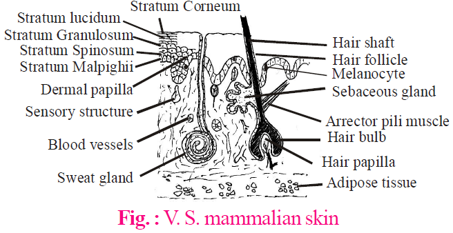
DERMIS
- It is differentiated into outer papillary layer and inner reticular layer. The papillary layer is compact with finger like projections, the papillae, whereas the reticular layer consists of loose connective tissue which merges with the subcutaneous fat.
- Below the dermis, there is a thin layer of fat, called Panniculus adiposus.
- The leather in mammals is produced from dermis part of the skin.
- Sensory Structures - Most of the sensory structures in the skin are ‘encapsulated dendrites’ and are embedded in the dermis. They all are ectodermal in origin.
- Meissner’s Corpuscles - They are sensory for touch (Thigmoreceptors).
- Merkel’s Discs - They are also sensory for touch or soft pressure.
- Pacinian Corpuscles - They are sensory for pressure (Baroreceptors)
- Krause end bulbs - They are sensory for cold (Frigidoreceptors).
- End bulbs of Ruffini - They are sensory for warmth (Caloreceptors)
- Free nerve endings - They are sensory for pain (Algesireceptors)
Due to a variety of functions, e.g., protection, temperature regulation, sensory, minimizing water loss etc., the skin is called ‘Jack of all trades’.
COCKROACH
- Cockroaches belong to class insecta of the phylum arthropoda.
- Bright yellow, red and green coloured cockroaches have also been reported in tropical regions. They are terrestrial, nocturnal, omnivores that live in damp places throughout the world.
- Systematic position of cockroach
Phylum – Arthropoda
Class – Insecta
Subclass – Pterygota
Order – Orthoptera
Genus – Periplaneta
Species – Americana
- Cockroaches (the name derived from the German word 'Cucaracha') are found in places where there is a warmth, dampness and plenty of organic food to devour.
- During day time, they remain inactive and hiding and during the night, they show much activity and run here and there in search of food showing their nocturnal habit. Their narrow and flattened body is adapted to slip into narrow creeks and crevices.
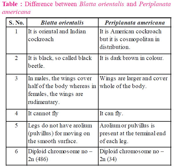
- Four species of cockroaches found in India are
- Blattella germanica (the German or Croton cockroach)
- Blatta orientalis (the Oriental or Indian Cockroach) : It is also called black beetle.
- Periplanata americana (the American cockroach or ship cockroach)
- Periplanata australasiae (the Australian cockroach)
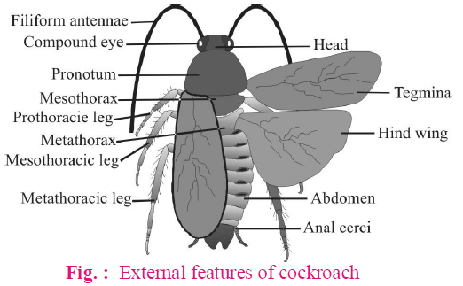
MORPHOLOGY
- The body of a cockroach is narrow, elongated and bilaterally symmetrical.
- Cockroach shows metameric segmentation.
- The adult cockroach measures 4 to 5 cms in length and 2 cm in width. Periplaneta americana is the biggest cockroach.
- The adult cockroach is reddish brown in colour. The nymph is white in colour.
The body of a cockroach is divisible into three parts – head, thorax and abdomen.
HEAD
- Head of cockroach is made by the fusion of 6 embryonic segments. It is triangular and pear shaped.
- Head is covered by an exoskeleton called head capsule. It is made by six sclerites.
- On the back of the head, an opening is present called occipital foramen. On its dorsal side, a small sclerite is present. It is called occipital.
- Head shows two compound eyes which are present on dorso-lateral sides. They are considered as the appendages of first head segment. These compound eyes are kidney shaped.
- The top of the head between the two compound eyes is called vertex.
- The six sclerites of head capsules are–
- Epicranial plates : Vertex is covered by 2 epicranial plates in the nymph stage. The two epicranial plates are separated by 'I' shaped suture.
- Frons : In front of vertex, a broad plate frons is present.
- Clypeus : Anterior to frons, there is a rectangular clypeus plate. Labrum is connected to clypeus.
- Genae plates : On the lateral sides of the head, two cheek sclerites are present. They are called genae.
- On the head, internal to the base of antennae, 2 white spots are present. They are called fenestra (or) ocellar spots. They work as photoreceptors.
- Near the ocellar spots on the head, two antennal sockets are present. They contain antennae. Each antenna shows a basal segment (called scape), second segment (called pedicle) and many segmented flagellum.
- Antennae are olfactory and tangoreceptors. Cockroach locates food with the help of antennae. Antennae are considered as the appendages of second segment of head.
- At the anterior end of the head, mouth parts are present.
THORAX
- Thorax is the middle part of the body.
- Thorax consists of three parts – prothorax, mesothorax and metathorax. The head is connected with thorax by a short extension of the prothorax known as the neck.
- Thorax bears 3 pairs of legs on the ventral side and two pairs of wings on the dorsolateral side.
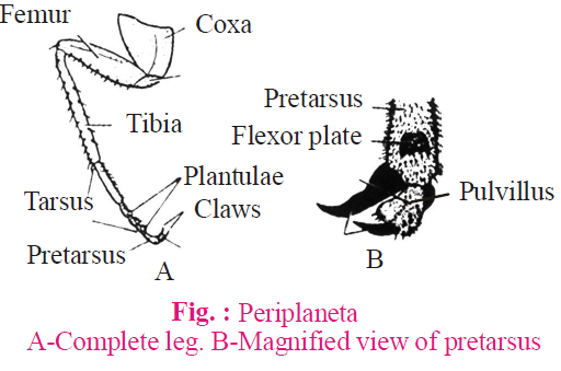
- Each leg is made by 5 podomeres –
- The first podomere is coxa. It is broad and attached to the thorax. It is the most swollen segment.
- The second podomere is small triangular trochanter. It gives viability for movement of leg. Trochanter is freely movable on coxa but fixed to the femur.
- The third podomere is femur. Femur is the strongest part of the leg. It shows bristles.
- The fourth podomere is tibia. Tibia is the longest part of the leg. It also bears bristles.
- The fifth podomere is tarsus. Tarsus contains 5 tarsomeres. The tarsomeres have plantulae on their ventral side. The last tarsomere is known as pretarsus which terminally contains a pair of claws. Between the two claws, a soft hairy pad is present called arolium or pulvillus. Arolium and plantulae serve as organs of attachment and provide a firm grip on smooth surfaces. When arolium and plantae are removed, cockroach cannot move on smooth surface.
- The first pair of wings arises from mesothorax and the second pair from metathorax. Forewings (mesothoracic), called tegmina or elytra, are opaque dark and leathery and cover the hind wings when at rest. The hind wings are transparent, membranous and are used in flight. They are folded like a fan and covered by elytra while at rest.
ABDOMEN
- Abdomen is broader than thorax and consists of 10 segments while embryo has 11 segments.
- 10th segment in both sexes bears a pair of small, filamentous and sensory anal cerci. Each anal cercus is made of 15 segments.
In male, in addition of anal cerci, 9th sternum bears anal style (unjointed thread like) which are absent in females.
- In female cockroach, abdomen is broader than in male.
BODY WALL
- Body wall of cockroach shows 3 parts– cuticle, epidermis and basement membrane.
- Cuticle is divisible into epi, exo and endo cuticular regions.
- Epicuticle : Epicuticle is the outermost layer of the cuticle. It is made by lipids and proteins. It is impermeable to water but permeable to CO2.
- Exocuticle : Exocuticle is made by thick hard and laminated chitin and phenols.
- Endocuticle : Endocuticle is made by proteins and soft chitin.
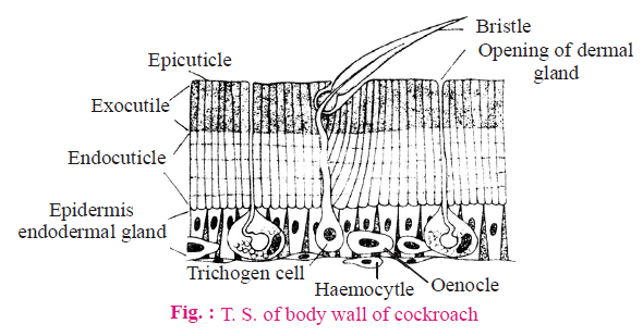
- The body of a cockroach is covered by chitinous exoskeleton. It is in the form of sclerites. The dorsal sclerite is called tergal plate. The ventral sclerite is called sternal plate.
- Two sclerites are connected by arthrodial membrane. This membrane is made by epicuticle and endocuticle, hence it is soft. It allows the movement of sclerites.
- Epidermis is made by a single layer of columnar cells. It shows supporting basal cells, trichogen cells, oenocytes etc. Trichogen cells give rise to movable bristles. Oenocytes produce cuticulin (wax). They also store glycogen, fats etc.
- Basement membrane forms a supporting layer of epidermis. It consists of an amorphous granular material (mucopolysaccharides)
- Functions of the body wall
- It covers and protects internal organs.
- It prevents the loss of water.
- It allows the loss of CO2 by diffusion.
- It shows sensory outgrowths.
- Outgrowths of cuticle serve as organs for locomotion and copulation.
- Body wall also provides a surface for the attachment of muscles.
- Male cockroach has longer antennae than females.
DIGESTIVE SYSTEM
Digestive system includes mouthparts, a long alimentary canal and a pair of salivary gland.
MOUTHPARTS
- Mouthparts of cockroaches are mandibulate type or cutting and chewing type.
- Mouthparts consist of labrum (upper lip), labium (lower lip), maxillae (segmented and resemble a leg), mandibles and hypopharynx (tongue).
- The main structures and well developed part of mastication (chewing) are mandibles which are short with teeth.
- The mandibles work as jaws and are used for crushing and cutting the food materials.
ALIMENTARY CANAL
- Alimentary canal is divided into three regions – foregut, midgut and hindgut.
- Foregut includes mouth cavity, pharynx, oesophagus, crop & gizzard.
- Mouth cavity is surrounded by mouth parts. It is divided into two parts – salivarium (into which common salivary duct opens) and cibarium (which leads towards mouth as narrow passage) by hypopharynx.
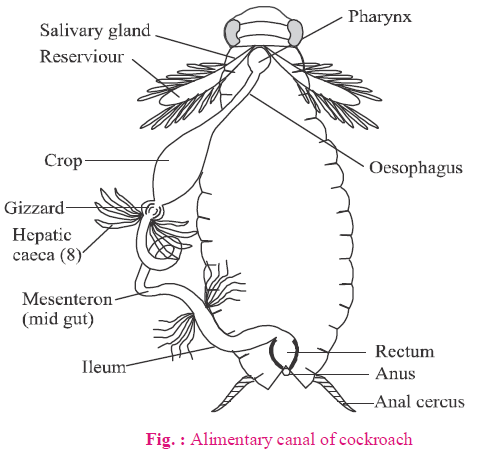
- Crop is the largest part of foregut & serve as a reservoir for storing food.
- The crop is followed by gizzard or proventriculus. It has an outer layer of thick circular muscles and thick inner cuticle forming six highly chitinous plate called teeth. Gizzard helps in grinding the food particles.
- The main part of gizzard is called armarium, anterior part with teeth (which masticate the food) and pulvilli (whose bristles form a strainer which allow only fine food particles to pass through).
- Stomodeal valve is posterior narrow and tubular part of gizzard which projects into the midgut to prevent backflow of food.
- A ring of 6-8 blind tubules called hepatic or gastric caeca is present at the junction of foregut and midgut, which secrete digestive juice.
- Midgut (mesenteron or ventriculus) is short, tubular lined with glandular endoderm.
- The anterior part of midgut consists of 4 parts of hepatic caeca.
- The lining of midgut secretes peritrophic membrane which protects the soft lining of midgut from the injury by spiniferus food. It is a temporary membrane and is removed after the food passes into the hindgut.
- Midgut forms the true stomach serving mainly for digestion and absorption.
- At the junction of midgut and hindgut is present another ring of 100-150 yellow coloured thin filamentous Malphigian tubules. They help in removal of excretory products from haemolymph. The hindgut is broader than midgut and is differentiated into ileum, colon and rectum. The rectum opens out through anus.
DIGESTIVE GLAND
- Digestive glands includes one pair of salivary gland, each is formed of two parts– sac like reservoir or (receptacle) and a bipartite glandular part.
- The digestive enzymes of saliva are mainly zymase and amylase.
- Digestion is intercellular in cockroach.
- Absorption of digested food takes place in mesenteron.
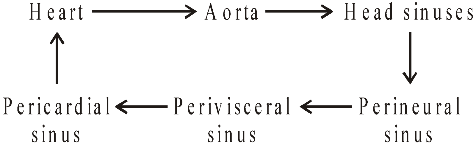
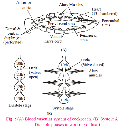
CIRCULATORY SYSTEM
- Circulatory system of cockroach is an open type. Blood vessels are poorly developed and open into space (haemocoel).
- Visceral organs located in the haemocoel are bathed in blood (hemolymph). The hemolymph is composed of colourless plasma and haemocytes.
- Heart of cockroach consists of elongated muscular tube lying along mid dorsal line of thorax and abdomen. It is differentiated into funnel shaped 13 chambers with ostia on either side. Blood from sinuses enter heart through ostia and is pumped anteriorly to sinuses again.
- The blood circulation is maintained by 13 pairs of wing-shaped involuntary alary muscles.
- Heart of cockroach is neurogenic (myogenic in frog, rabbits and man).
- In addition to the main heart, there are present very small accessory heart or pulsatile vesicles one at the base of each antenna located in the head, to pump the blood from the head sinuses to the antenna.
- The blood of cockroach is colourless due to the lack of respiratory pigment.
- All body tissues receives oxygen directly.
- The rate of heart beat in Periplaneta is 49 beats/minutes
- Blood circulation in cockroach is completed in 5-6 minutes.
The pathway follow as :
RESPIRATORY SYSTEM
- The respiratory system of cockroach consists of a network of trachea, that open through 10 pairs of small holes called spiracles (two pairs thoracic and eight pairs of abdominal) present on the lateral side of the body. Thin branching tubes (tracheal tubes subdivided into tracheoles) carry oxygen from the air to all the parts
- The opening of the spiracles is regulated by the sphincters.
- Exchange of gases takes place at the tracheoles by diffusion.
- The trachea is lined with spiral thickening of cuticle called intima or taenidia which prevents the tracheal tubes from collapsing (trachea of rabbit is also non collapsible).
- Ventilation of tracheal system is by alternate contraction and relaxation of abdominal muscles (tergo-sternal muscles).
- Respiratory movement are coordinated and regulated by nerve centres in thoracic ganglia which are stimulated by low O2 and higher CO2 concentrations in tissue fluids.
Tracheal systems of respiration is also found in centipedes, millipedes, ticks and Peripatus.
EXCRETORY SYSTEM
- Excretion is performed by Malpighian tubules. Each tubule is lined by glandular and ciliated cells. They absorb nitrogenous waste products and convert them into uric acid which is excreted out through the hindgut. Therefore, this insect is called uricotelic.
In addition, the fat body, nephrocytes and uricose glands also help in excretion.
- Fat body of cockroach contains mainly four types of cells, viz., trophocytes, mycetocytes, oenocytes and urate cells.
- The trophocytes are most numerous containing reserve food in the form of fats, glycogen and proteins.
- Mycetocytes contain symbiotic bacteria which help in synthesis of some amino acids, vitamins and of glycogen from glucose.
- Oenocytes are supposed to help intermediary metabolism at times of ecdysis. It secretes wax which covers the cuticle of cockroach.
- Urate cells absorb nitrogenous waste products from haemolymph and synthesize uric acid from these for storage (storage excretion).
- The fat body of cockroach is functionally analogous to liver of vertebrates.
- Uricose gland are long, blind tubules present at the periphery of mushroom gland in the male cockroach.
- These tubules store uric acid and discharge it over the spermatophore during copulation.
- Uricose gland serves as storage excretory organs between matings and as active excretory organs during copulation.
NERVOUS SYSTEM
- The nervous system of cockroach consists of a series of fused, segmentally arranged ganglia (total 9) joined by paired longitudinal connectives on the ventral side. Three ganglia lie in the thorax, and six in the abdomen.
- The nervous system of the cockroach is spread throughout the body.
- The head holds a bit of a nervous system while the rest is situated along the ventral (belly-side) part of its body.
- Cockroach has a well developed nervous system with central, peripheral and sympathetic system.
- Central nervous system consists of cerebral or supraesophageal ganglion (brain), suboesophageal ganglion, paired circumesophageal connectives and double ventral nerve cord (with three thoracic and six abdominal compound segmental ganglia).
- Sense organs in cockroach are – photoreceptors (compound and simple eye), thigmoreceptors (antennae), chemoreceptors (on maxillary and labial palps, labium and hypopharynx) and auditory receptors on anal cerci.
- Each compound eye of cockroach is composed of about 2000 visual units called ommatidia.
- Each ommatidium is composed of a cuticular lens, two corneagen cells, a crystalline cone surrounded by four cone cells, a rhabdome surrounded by seven reticular cells and a basement membrane.
- There are two types of vision in insects – mosaic vision (or apposition image) during the day time and superposition (or dull image) in dim light.
- But in cockroach, pigment sheath of ommatidia is non-contractile so capable of only mosaic vision even during the night.
REPRODUCTIVE SYSTEM
- Cockroaches are dioecious and both sexes have well developed reproductive organs.
- Male organs consist of testes, vasa deferentia, ejaculatory duct, mushroom or utricular gland, phallic or conglobate gland and male gonapophysis.
- Testes of cockroach are located in the abdominal segments 4, 5 and 6.
- From each testis arises, a thin vas deferens, which opens into ejaculatory duct through seminal vesicle.
- The ejaculatory duct opens into male gonopore situated ventral to anus.
- A characteristic mushroom shaped gland is present in the 6th-7th abdominal segments which functions as an accessory reproductive glands.
- Mushroom gland consists of two types of tubules, the long slender tubules, the utriculi majores or peripheral tubules; and short tubules, the utriculi breviores, making up of the major part of the gland.
- The external genitalia are represented by male gonapophysis or phallomere (chitinous asymmetrical structures, surrounding the male gonopore).
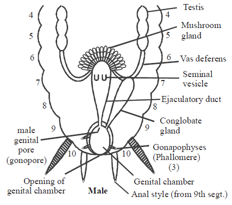
- The sperms are stored in the seminal vesicles and are glued together in the form of bundles called spermatophores which are discharged during copulation.
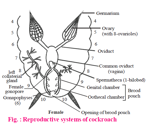
- The female reproductive system consists of two large ovaries, lying laterally in the 2nd – 6th abdominal segments.
- Each ovary is formed of a group of eight ovarian tubules or ovarioles, containing a chain of developing ova.
- Oviducts of each ovary unite into a single median oviduct (also called vagina) which opens into the genital chamber. A pair of spermatheca is present in the 6th segment which opens into the genital chamber and receives sperms from spermatophores. Their fertilized eggs are encased in capsules called ootheca. Ootheca is a dark reddish to blackish brown capsule, about 3/8" (8 mm) long. They are dropped or glued to a suitable surface, usually in a crack or crevice of high relative humidity near a food source.
- On an average, females produce 9-10 ootheca, each containing 14-16 eggs.
- The development of P. americana is paurometabolous, meaning there is development through nymphal stage. The nymphs look very much like adults. The nymph grows by moulting about 13 times to reach the adult form. The next to last nymphal stage has wing pads but only adult cockroaches have wings.
- Metamorphosis is regulated by two hormones ecdysone (secreted by prothoracic glands) and juvenile hormone or neotenin (secreted by corpora allata).
EARTHWORM
Taxonomic position
Phylum - Annelida
Class - Oligochaeta
Order - Opisthopora/Haplotaxida
Genus - Pheretima
Species - posthuma
- It is metamerically segmented with 100-120 segments (metameres), each separated by inter segmental groove. The number of segments remain fixed throughout life, i.e. neither new segments are added, nor the old ones are removed.
- It is burrowing (fossorial).
- The skin is brown due to the presence of Porphyrin pigment which is derived from chlorophyll, taken by the young animal with the soil.
- The dorsal side of the body can be distinguished by the presence of a prominent dorsal blood vessel. The ventral side contains two pairs of prominent genital papillae, one pair in each segment number, 17th and 19th.
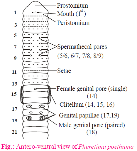
- The first segment, called Peristomium, bears mouth on the ventral side. The hood like projection of peristomium is called prostomium.
- The male genital apertures (gonopores) are two (one pair), present on ventral side of 18th segment.
- Female genital aperture is single and is present in mid-ventral side of 14th segment.
- Spermathecal pores are four pairs and are present in intersegmental grooves of segments 5/6, 6/7, 7/8 and 8/9.
- The anal opening is present in the last segment.
- The body of the earthworm can be differentiated into 3 regions –
- Pre-clitellar region (1st to 13th segments)
- Clitellar region (14th, 15th and 16th segments).
- Post-clitellar region (17th to last segment).
(As the number of total segments ranges from 100 to 120, the number in post-clitellar region may be different in different earthworms. The number of segments in preclitellar region and clitellar region is fixed.)
SEPTAE
- They are the partitions between adjacent segments in intersegmental grooves.
- The septae are absent from segments 1st to 4th. The presence of continuous-coelomic fluid in these segments make them turgid, to help in burrowing.
- Septae are also absent between 9th and 10th segments.
- In rest of the segments ( up to 14th ), the septae are non-porous.
- From 14th / 15th segments, all the septae are porous and coelomic fluid, acting as hydrostatic skeleton, is interconnected.
BODY AND COELOM
- Body wall consists of 4 layers, i.e. cuticle, epidermis, circular muscles and longitudinal muscles.
- Earthworm is Eucoelomate, having schizocoel, lined with somatic/parietal mesoderm on the outside and visceral/splanchnic mesoderm in the inner side.
- Coelom contains coelomic fluid, acting as hydrostatic skeleton. This fluid oozes out from the body through dorsal pores in intersegmental grooves, one per segment, from 12/13th to the last of the body.
LOCOMOTION
- It is carried out by body wall musculature (circular muscles and longitudinal muscles).
- The setae are chitinous structure present in setigerous sac and are produced from formative cells. They are embedded in the body wall and can move with the help of protractor and retractor muscles.
- Coelomic fluid, forming hydrostatic skeleton, and the setae help in locomotion.
- This process of locomotion, called ‘Thinning and thickening process’ was described by Gray and Lissman.
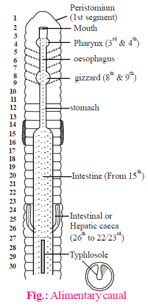
NUTRITION
- Earthworm is soilivorous/detritivorous or omnivorous.
- The alimentary canal is complete and highly specialized. It consists of the following parts –
- Mouth and buccal cavity
- Pharynx
- Oesophagus
- Gizzard
- Stomach
- Intestine
- Rectum
- Anus
- The pharynx (segments 3rd and 4th) is suctorial and is differentiated into two chambers, dorsal chamber and ventral chamber. Pharyngeal gland or salivary gland is present in the dorsal side of pharynx. Salivary glands secrete saliva into the dorsal chamber. The chromophil cells of pharyngeal gland/salivary gland are analogous to the chief cells of vertebrate stomach, as they secrete proteolytic enzymes, like pepsin, which digest food in acidic medium in the presence of humus acid of the soil.
- Gizzard (segment 8th – 9th) is a masticatory organ and helps in cutting down the food material due to muscular movements. It has cuticular lining. The longitudinal muscles in gizzard are absent.
- Stomach extends from 9th to 14th segment. In earthworms, other than Pheretima posthuma, there are calciferous glands in the stomach to neutralize the acidic medium.
- Intestine is the longest and widest part of the alimentary canal and extends from 15th segment to the last. It is meant for digestion and absorption of the food material. One pair of intestinal caeca arise from 26th segment and extend forward up to 22/23rd segment. These caeca, also called hepatic caeca, secrete amylase for carbohydrate (starch) digestion. The other enzymes secreted from intestinal lining are cellulase, chitinase, lipase, trypsin etc.
- Intestine is differentiated into 3-parts, namely -
- Pre-typhlosolar region (15th to 26th segment)
- Typhlosolar region (27th to leaving last 25 segments)
- Post-typhlosolar region (last 25 segments)
- The post typhlosolar region (last 25 segments), also known as rectum, is for the storage of undigested waste, which is removed from the gut in the form of ball-shaped castings.
RESPIRATION
- Earthworm respires through moist skin (cutaneous respiration). The skin is kept moist by coelomic fluid oozing out through dorsal pores.
- Because of the cylindrical shape the ‘surface area to volume ratio’ is higher in earthworm.
- There is no specialized respiratory or breathing structure in the body.
EXCRETORY SYSTEM
The excretory structures in earthworm are nephridia (metanephridia). They are ectodermal in origin and are of 3-types:
- Integumentary Nephridia
- Pharyngeal Nephridia
- Septal Nephridia
INTEGUMENTARY NEPHRIDIA
- They are present in the skin/integument of segments number 3rd to last.
- They are maximum in clitellar region (14, 15 and 16th segments ). The term ‘Forest of nephridia’ is used for these nephridia in clitellar region, as their number ranges from 2000-2500 per segment.
- The number of these nephridia in other segments is 200-250 per segment.
- They are the simplest nephridia and do not have nephrostome.
- The excretory matter is taken from blood and is poured outside the body through nephridiopores. That is why such nephridia are called Exonephric nephridia or Exteronephric nephridia.
PHARYNGEAL NEPHRIDIA
- They are present near pharynx in segments number 4th, 5th and 6th only.
- There is one pair of tuft/bunch per segment. Each tuft has about 100 nephridia (200 nephridia per segment).
- Out of 3-types of nephridia, the pharyngeal nephridia are minimum in number.
- They too lack nephrostome and are, therefore, simple in structure.
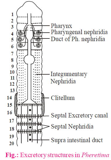
- Like integumentary nephridia, they also take excretory matter from the blood.
- These nephridia pour the excretory matter into pharynx through nephridial ducts.
- Such nephridia are called Endonephric nephridia or Enteronephric nephridia.
SEPTAL NEPHRIDIA
- They are attached to intersegmental septae, from 15/16th septa, to the last.
- They are called typical nephridia or Holonephridia as they are complete and contain nephrostome.
- The number of these nephridia ranges from 80-100 per segment.
- They collect excretory matter from blood as well as coelom, from latter through nephrostome.
- The excretory matter is poured through septal excretory canal and supra intestinal duct, into intestine.
- Such nephridia are also known as Endonephric nephridia or Enteronephric nephridia since the excretory matter goes outside through the gut. (Both pharyngeal and septal nephridia pour the excretory product into gut for water absorption, and are thus adapted for water conservation.)
- Each septal nephridium consists of following parts–
- Nephrostome
- Twisted lobe (proximal and distal limb)
- Straight lobe
- Terminal duct
- The route of excretory product through septal nephridia is –
Nephrostome → proximal limb → distal limb → straight lobe → distal limb → proximal limb → terminal duct
- The excretory product in earthworm is urea (ureotelic). In rains, due to plenty of water, the major excretory product becomes ammonia.
- The chloragogen cells, present in the visceral mesoderm of gut and coelomic fluid of the body, synthesize urea and other excretory materials like chloragogue formed by the metabolism of haem. The chloragogen cells also store reserve food material, and are, therefore, comparable(analogous) to vertebrate liver.
BLOOD VASCULAR SYSTEM (BVS)
- BVS in earthworm is closed type.
- Haemoglobin is dissolved in plasma and RBCs are absent. WBCs or phagocytic cells are present.
- The blood is synthesized in blood glands in segments 4th, 5th and 6th
- The pharyngeal nephridia and blood glands occur together in the same segments, i.e., 4th, 5th and 6th.
- The lymph glands which produce phagocytic cells and release them into coelomic fluid, are 1 pair per segment (from 26th to last). They are dorso-lateral in position.
- The blood vascular system in earthworm can be studied in 2-parts-
BVS IN FIRST 13-SEGMENTS
It consist of following prominent structures
- Dorsal blood vessel (DBV).
- Supra-oesophageal blood vessel (SOBV)
- Ventral blood vessel (VBV)
- Lateral Oesophageal blood vessel (LOBV)
- Hearts
- Anterior loops
(1) Dorsal Blood Vessel
- It is single or unpaired and prominent blood vessels in the mid-dorsal side of the body.
- It is distributive in nature and supplies blood to the anterior part of the gut, i.e. buccal cavity, pharynx, oesophagus, stomach and gizzard.
- It has one pair of valve per segment and is comparable to the veins of vertebrates or heart of cockroach.
- The direction of blood, flow is from posterior to anterior side.
- It is connected to ventral blood vessel through two pairs of false and two pairs of true hearts.
(2) Supra-Oesophageal Blood Vessel
- It is also single or unpaired blood vessel, present in segments number 9th to 13th only.
- It is collective in nature and collects the blood from anterior part of alimentary canal.
- The valves are absent in this blood vessel.
- It receives blood from LOBV through anterior loops.
(3) Ventral Blood Vessel
- It is also single or unpaired vessel present in the mid-ventral side of the body.
- It is distributive in nature.
- It supplies blood to nephridia, body-wall, septae and reproductive organs.
- The direction of blood flow is from anterior to posterior side.
- The valves are absent.
- It receives blood from dorsal blood vessel through false and true hearts.
(4) Lateral Oesophageal Blood Vessel
- It is paired and lateral in position.
- It is collective in nature, and collects the blood from skin, excretory organs, reproductive organs etc.
- The direction of blood flow is from anterior to posterior side.
- Valves are absent.
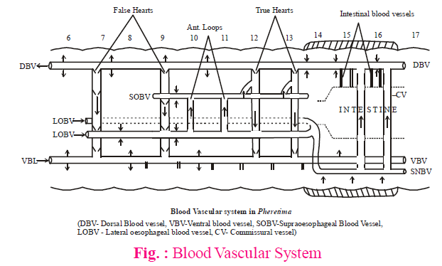
(5) Hearts
- There are 4-pairs of hearts (8 in number) in the earthworm.
- They are of 2-types –
- Lateral hearts
- They are also known as false hearts.
- They are 2-pairs in number, 1 pair each in segments, 7th and 9th.
- They connect dorsal blood vessel to ventral blood vessel.
- The direction of blood blow is from dorsal to ventral side.
- There are 4-pairs of valves per lateral heart.
- Lateral esophageal hearts
- They are also known as true hearts.
- They are also 2-pairs in number, 1 pair each in segments, 12th and 13th.
- They connect dorsal blood vessel and supra oesophageal blood vessel to ventral blood vessel.
- Direction of blood flow is from dorsal to ventral side.
- Each heart has 3-pair of valves, 1 pair at each opening of dorsal/ventral and supra oesophageal blood vessels.
(6) Anterior loops
- There are 2 pairs of anterior loops (4 in number).
- They are non-pulsating in nature and are present in segments, 10th and 11th.
- They connect supra-oesophageal blood vessel to lateral esophageal blood vessel.
- The direction of blood flow is from ventral to dorsal side (LOBV to SOBV)
- Valves are absent.
BVS AFTER 13-SEGMENTS
The blood vascular system from 14th to the last segment consists of the following important vessels.
- Dorsal blood vessel (DBV)
- Ventral blood vessel (VBV)
- Subneural blood vessel (SNBV)
- Commissural vessels
- Intestinal vessels
(1) Dorsal blood vessel
- After 13-segments, it becomes collective in nature and collects blood from the alimentary canal, excretory organs and reproductive organs. (Since DBV collects blood from about 100 segments (14th to last) it is mainly collective in nature).
- The supra-oesophageal blood vessel, collecting blood from anterior part of the gut, is absent after 13th segment.
(2) Ventral blood vessel
It is the same throughout the body.
(3) Sub-neural blood vessel
- Two LOBV unite after 13th segment to form single blood vessel which runs posteriorly below the ventral nerve cord, and is so called sub-neural blood vessel.
- The sub-neural blood vessel is connected to dorsal blood vessel through commissural vessels.
(4) Commissural vessels
- They are 2 (1 pair) per segment and are present after 15th segment to the last.
- They connect dorsal blood vessel to sub-neural blood vessel.
- The blood flow is from ventral to dorsal side.
(5) Intestinal blood vessels
- They are 2-pairs per segment.
- They are collective in nature and pour the blood into dorsal blood vessel.
NERVOUS SYSTEM
Earthworm contains central nervous system (CNS), peripheral nervous system (PNS) as well as autonomous nervous system (ANS)
- The central nervous system consists of a nerve ring and a paired ventral nerve cord.
- The nerve ring is formed by supra-pharyngeal ganglia, circum-pharyngeal connectives and sub-pharyngeal ganglia. The ring is present around pharynx.
- Supra pharyngeal ganglia form the brain of the earthworm.
- The ventral nerve cord is paired but fused to appear as single, and is enclosed in a common sheath. It is ganglionated, having 1-pair of ganglia (fused) per segment.
- Above ventral nerve cord there are 4-giant nerve fibers, having greater diameter, for conducting the impulses at a faster pace.
SENSORY ORGANS
- The sensory structures in skin are thigmoreceptors.
- The dorsal side of anterior segments, particularly peristomium and prostomium, contains photoreceptor cells. Each photoreceptor cell has a lens like structure, called phaosome. These cells do not form the image but detect the presence of light.
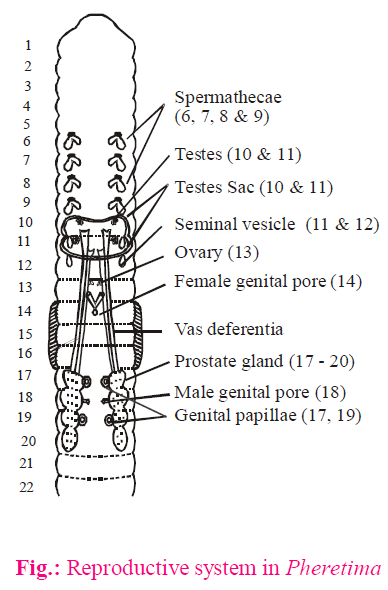
REPRODUCTIVE SYSTEM
- Earthworm is bisexual (monoecious) or hermaphrodite.
- Both male and female reproductive systems are present in antero-ventral side of the body.
MALE REPRODUCTIVE SYSTEM
- It consists of the following parts -
- Testes - 2-pairs (10th and 11th segments)
- Testes sac - 2 (10th and 11th segments)
- Vas deferens - 2-pairs.
- Seminal vesicle - 2-pairs (11th and 12th segments)
- Prostate gland - 1-pair (17th to 20th segments)
Genital aperture - 1 pair (18th segment)
- Testes are present in the testes sac. They produce spermatocytes which enter into seminal vesicles. The maturation of sperms occurs in seminal vesicles (not testes).
- Sperms from seminal vesicle pass into vas deferens through spermatic funnel.
- The sperms with the secretion of prostate gland are released through male genital apertures as spermatic fluid.
- The accessory glands, for adhesive secretion, open into genital papillae in segments number 17th and 19th
FEMALE REPRODUCTIVE SYSTEM
- It consists of the following parts -
- Ovaries - 1-pair (13th segment)
- Oviducts - 1-pair
genital aperture - Single (14th segment)
- Spermatheca - 4 pairs (6th, 7th, 8th and 9th segments)
- Each ovary consists of a number of ovarioles, and is attached to the septa of 12th/13th segment.
- The eggs pass through oviducal funnel and oviduct, and are released through single genital aperture in 14th segment.
- The spermathecae are differentiated into outer ampulla and inner diverticulum, and open in intersegmental groove in 5th/6th , 6th/7th, 7th/8th and 8th/9th segments. In earthworm (Pheretima) the sperms, of the other earthworm, are stored in the diverticulum part of the spermathecae.
BREEDING AND FERTILIZATION
- It occurs before dawn, during the rainy season.
- Earthworm is protandrous and self fertilization is absent.
- Fertilization is cross and reciprocal.
- The two earthworms get attached in the antero-ventral side with the help of genital papillae. The male genital apertures of one earthworm face the first pair of spermathecal opening of 5th/6th segment of the other earthworm.
- During mating, the sperms of one earthworm are transferred into the diverticulum of spermathecae of the other earthworm, and vice-versa.
- The eggs are released through female genital aperture in 14th segment (anterior clitellar region).
- The epithelium of clitellum secretes cocoon. This cocoon contains unfertilized eggs.

- Now, earthworm wriggles backwardly and comes out of the cocoon through its anterior end. When the cocoon comes around the openings of spermathecae, the sperms are released over the eggs and the eggs get fertilized in the cocoon. Thus, fertilization is external but in cocoon.
- The epidermal glands also secrete albumin which is stored in the cocoon as a reserve food material for developing embryo.
DEVELOPMENT
- In earthworm, the development is direct, i.e. without larva formation.
- Out of 2 - 20, usually 4 - embryos are matured in the cocoon, and the rest are used as food.
ECONOMIC IMPORTANCE
- Earliest use of earthworm is in Unani and Ayurvedic medicines for the treatment of gout.
- Earthworms are also used as bait for fishing.
- They are natural ploughmen and make the soil porous.
- They enrich the soil (increase the fertility) by adding excretory and faecal matter.
- The ‘vermicompost’ is in high demand, these days.
FROG
Taxonomic position
Phylum - Chordata
Class - Amphibia
Order - Anura
Genus - Rana
Species - Tigrina
- Rana tigrina is commonly called as Indian Bull-frog.
- It is amphibious, and can be found in ponds/shallow water bodies, and in terrestrial conditions. Its close relative, Toad (Bufo melanostictus) is largely terrestrial.
- It is cold blooded or Poikilothermic and shows summer-sleep (Aestivation) and winter-sleep (Hibernation) in extreme temperatures.
- Its body colouration shows camouflage. (Its colour changing ability is called Metachrosis)
- It is insectivorous/carnivorous.
EXTERNAL MORPHOLOGY
- The body of the frog is streamlined, well adapted for swimming, leaping and burrowing.
- It skins is slimy, moist without exoskeleton or scales. Skin contains mucus and poison glands, the latter are concentrated on shoulders. The chromatophores in the skin help in camouflage.
- Eyes are dorso-laterally placed on head. Frog has 3-eyelids, i.e. upper eye-lid, lower eye-lid and third eye-lid (Nictitating membrane), the layer covering the eye-ball during swimming.
- External ear is absent in frog. The tympanum/eardrum is present near the eyes.
- The body of the frog is divisible into head and trunk. The neck is absent. The trunk has 1-pair of shorter fore limbs and 1-pair of longer hind limbs. Each hand has 4-fingers (digits) whereas each foot has 5-toes (digits). (The thumb, pollex, or the first finger in hand is absent in frog.)
DIGESTIVE SYSTEM
- Due to carnivorous nature, the alimentary canal of frog is short. The posterior most part of the gut is cloaca. Urine also passes through cloacal opening.
- Due to absence of Uvula, the buccal cavity and pharynx are not differentiated. The bucco-pharynx contains numerous maxillary teeth. The teeth in frog are Homodont (similar type), Acrodont (fused with the Jaw-bones) and Polyphyodont (many sets of teeth). The teeth are, however, absent in lower-jaw. Besides upper-jaw, the teeth are also present in vomer bone of olfactory capsule, in the roof of bucco-pharynx. The teeth in frog are not used for cutting the food material, but are used for preventing the escape of the prey. The tongue in frog is bifid at the tip and is free behind. It is used for capturing prey (insects).
RESPIRATORY SYSTEM
- Frog respires through skin (Cutaneous respiration), bucco-pharyngeal lining and through the lungs (Pulmonary respiration). During aestivation and hibernation, the only mode of respiration is cutaneous.
- The lungs are hollow, non-lobular and positive pressure type. The pulmonary respiration has 3-phases, i.e. Aspiration, Inspiration and Expiration. During aspiration, sternohyoid muscles of lower-jaw contract and air through nostrils is filled into bucco-pharynx. During inspiration, the lower-jaw is raised (due to contraction of petrohyal muscles) and nostrils are closed. The air is forced into the lungs (+ve pressure lungs). In expiration, the air comes out of the body through nostrils.
BLOOD VASCULAR SYSTEM
- In frog, BVS is closed type and has double circulation, though not as efficient as in birds and mammals.
- The heart is 3-chambered (2-atria and 1-ventricle). The dorsal side of the heart has a triangular structure, called Sinus venosus which receives deoxygenated blood from
2-precavals and 1-postcaval. It pours the blood into the right atrium. On the ventral side of the heart, there is a prominent structure, called Conus arteriosus which arises from the ventricle. Conus arteriosus is differentiated into proximal Pylangium (bulbus arteriosus) and distal Synangium (ventral aorta). The Pylangium contains cardiac muscles and is the part of the heart. It also contains a fold-like Spiral valve which divides it into Rt- ventral chamber, Cavum aorticum, and Lt-dorsal chamber, Cavum Pulmocutaneous. Each branch of ventral aorta contains 3-arches i.e. Pulmocutaneous (Dorsal), Systemic (middle) and common carotid (ventral). - The SA node is present in the wall of sinus venosus and not in the right atrium, as in mammals.
- As there is a single ventricle (interventricular septum absent), the bicuspid and tricuspid valves are not differentiated and blood of the 2-atria is mixed in ventricle.
- The AV node and Bundle of His are absent in frog.
- There are 2-types of portal systems in the body of a frog, i.e. Hepatic portal system (between gut and liver) and Renal portal system (between posterior abdominal parts and kidney).

(BA - Bulbous arteriosus or Pylangium, VA - Ventral aorta or Synangium)
RENAL PORTAL SYSTEM
- The femoral vein, arising from outer thigh region, after entering abdomen, bifurcates to form outer renal portal vein and inner pelvic vein.
- The renal portal vein terminates into kidney whereas the pelvic veins of two sides join to form Anterior Abdominal Vein. The anterior abdominal vein finally terminates into liver.
- The Sciatic vein that arises from inner thigh region joins renal portal vein and pours its blood into that.
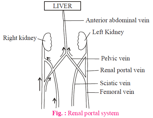
NERVOUS SYSTEM
- Frog has all 3-types of nervous system, i.e. CNS (brain and spinal cord), PNS (Cranial nerves and spinal nerves) and ANS (SNS and PSNS).
- The cranial and spinal nerves in frog are 10-pairs each. However, in Rana tigrina the number of spinal nerves is 9-pairs.
- In brain, the cerebral hemispheres are less developed than in mammals. The corpus callosum, connecting two cerebral hemisphere internally in mammals, is absent in frog.
- The midbrain consists of 2 optic lobes (Corpora bigemina). Both lobes are hollow with optocoel.
- Hindbrain consists of cerebellum and medulla oblongata. The pons is absent in frog.
SENSORY ORGANS
- The eye-ball in frog is cartilaginous. The colour vision is lacking due to absence of cones in the retina. In place of lacrimal and meibomian glands, there are Harderian glands in frog.
- The sensory part for hearing in the internal ear of frog is Pars basilaris & Pars lagena. The Cochlea and Organ of Corti are absent. The round window (Fenestra rotundus) between middle and internal ear is also absent.
- In olfactory capsule, the Septo-maxillary bone is the smallest bone of the frog’s body.
EXCRETORY SYSTEM
- The kidneys in frog are mesonephros. Such kidney is not differentiated into cortex and medulla.
- Each kidney has 4-5 pairs of renal artery and veins.
- The nephrons or uriniferous tubules are not differentiated into PCT, DCT and Henle’s loop.
- The major excretory product in frog is urea (Ureotelic).
REPRODUCTIVE SYSTEM
The sexual dimorphism in frog is prominent during the breeding season. The male frog differs from female frog in the following features.
- Male frog has nuptial pad at the base of the first finger (actually second finger, as pollex thumb is absent)
- Males have vocal sacs for amplification of sound.
- The colour pattern of the male is also different from the female body.
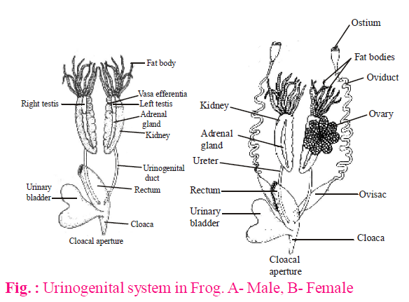
MALE REPRODUCTIVE SYSTEM
- The testes are attached to kidney through fold of peritoneum, called mesorchium.
- The sperms from testes pass through vasa-efferentia (10-12 in number) and are released into Bidder’s canal, a collecting duct of the kidney. It is because the vas-deferens is absent in frog.
- The mesonephric duct (or Wolffian duct) of kidney is, therefore, also known as urino-genital duct. Faecal matter, urine and sperms, all are released through cloacal opening.
FEMALE REPRODUCTIVE SYSTEM
The ovaries are also close to kidney, but unlike testes, are not functionally connected to it. The oviducts open separately into cloaca.
DEVELOPMENT
- The Frog is spontaneous or seasonal ovulator, i.e. the Ovulation occurs according to the season. (Rabbit is Induced or Reflex ovulator, i.e. copulation induces ovulation). The eggs (2000 - 4000) are laid in spawn.
- The eggs in frog are mesolecithal and telolecithal. The animal hemisphere of the egg is pigmented due to the presence of melanin and shows camouflage. The eggs are at secondary oocyte stage with one polar body. Each egg is covered by an inner non-living, vitelline membrane and outer jelly, the latter secreted from oviducts.
- The jelly is protective (shock absorber), distasteful, adhesive and non-conductor of heat. It is light in weight and keeps the eggs floating on water surface to ensure them sufficient oxygen and sunlight. The jelly also contains ‘Fertilizin’ for the attachment of sperms.
- The copulation (without copulatory organ in male) is false and is called Amplexus.
- The fertilization is external (in water). After fertilization, due to movement of melanin particles there develops a ‘Gray crescent’, opposite to the sperm entrance point. The 1st cleavage occurs through the middle of Gray crescent to establish bilateral symmetry.
- The cleavage is unequal holoblastic (first and second cleavages are, however, equal holoblastic).
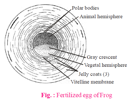
- The development is indirect. The larva is tadpole.
- The important stages of development are:
Zygote → Morula → Blastula → Gastrula → Neurula → Tail-bud stage → Gill bud-stage → External gill stage → Internal gill stage ( mature tadpole).
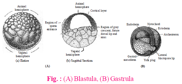
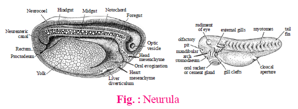
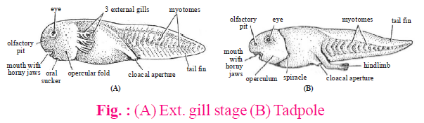
- Mature tadpole has pronephros kidney, cartilaginous skeleton and respires through skin and internal gills (4-pairs). The blood vascular system is fish like. The heart is 2-chambered. The alimentary canal is coiled like a spring. It is a herbivore. The forelimbs and hind limbs are developed, but the forelimbs are not visible due to the presence of operculum over gills. The excretory product is ammonia.
METAMORPHOSIS
- It occurs in the presence of thyroxine hormone. In deficiency of thyroxine or iodine, the tadpole increases in size and does not undergo metamorphosis.
- The emergence of fore limbs out of operculum is the first sign of metamorphosis.
- The nervous system is least affected and blood vascular system is most affected during metamorphosis.
- The important destructive/regressive changes in metamorphosis are –
- Shedding of horny jaws and teeth
- Resorption of tail by autolysis
- Disappearance of cloacal tube, lateral line organ, operculum, spiracle and internal gills.
ECONOMIC IMPORTANCE OF FROG
- It is used in laboratory for teaching and research.
- Being insectivorous, it protects the crops from insect-pests and saves the expenditure on insecticides.
- Its muscular legs are used as food/pickle.
- Froglets or tadpoles can be used as fish baits.
- Frog also forms an important link in ecosystem and maintains ecological balance.
Study Notes for NEET/AIIMS/JIPMER


