


CELL : THE UNIT OF LIFE
INTRODUCTION
- Just as a building is made up of bricks, the bodies of all plants and animals are made up of cells, i.e., all living beings show cellular organisation. Some organisms like Amoeba, Paramecium, bacteria, Chlamydomonas, etc. are made up of one cell (unicellular) only. There are a large number of other organisms like plants, animals, human beings etc.which are made up of millions of cells (multicellular).
- Cell is the structural and functional unit of all living beings.
- Robert Hooke (1665) discovered hollow cavities (empty boxes) like compartments in a very thin slice of cork (cell wall) with his crude microscope and named them as cellulae or cells.
- Anton Von Leeuwenhoek first saw and described a live cell.
- Robert Brown later discovered and named the nucleus in a cell. J. E. Purkinje, used the term protoplasm for the living substance present inside the cell.
- The invention of the microscope and its improvement leading to the electron microscope revealed all the structural details of the cell.
- Electron microscope : This was developed by Max Knoll and Ernst Ruska (1931) in Germany. It is a large sized instrument which has an internal vacuum, high voltage (50,000 – 1,00,000 volts), a cooling system, a fast beam of electrons (0.54 Å wavelength), a cathode filament of tungsten and electromagnetic lens (which has a coil of wire enclosed in soft iron casing) for focusing. It can magnify objects upto 2,00,000 times (now possible upto 2,50,000 – 4,00,000). The resolving power of electron microscope is 10 Å which is 100 times more than the light microscope. Study of living cells cannot be done through this microscope because of high voltage, which is required to operate it, as that can kill the living materials.
CELL THEORY
- The actual credit for cell theory goes to two scientists, a German Botanist M.J. Schleiden (1838) and a British Zoologist T. Schwann (1839). They gave the concept "all living organisms are composed of cells" and products of cells. Schleiden and Schwann together formulated the cell theory but this theory did not explain how new cells were formed.
- Viruses, viroids and prions are exceptions to the cell theory as they are obligate parasites (subcellular in nature).
- Rudolf Virchow (1855) first explained that cells divide and new cells are formed from pre-existing cells (Omnis cellula-e cellula). He modified the hypothesis of Schleiden and Schwann to give the cell theory a final shape. Cell theory states that–
- All living organisms are composed of cells and products of cells.
- All cells arise from pre-existing cells.
- Membrane bound cell organelles of the protoplasm do not survive along or outside the protoplasm.
- All cells have similar fundamental structure and metabolic reactions.
- Genetic information is stored as DNA in the chromosomes present in the nucleus.
AN OVERVIEW OF CELL
- Each cell has a dense membrane bound structure called nucleus which contains the chromosomes that in turn contain the genetic material, DNA.
- Cells that have membrane bound nuclei are called eukaryotic whereas cells that lack a membrane bound nucleus are prokaryotic.
- The eukaryotic cells have membrane bound distinct structures called organelles like the endoplasmic reticulum (ER), the golgi complex, lysosomes, mitochondria, microbodies and vacuoles. The prokaryotic cells lack such membrane bound organelles.
- Cells differ greatly in size, shape and activities.
For example, Mycoplasmas, the smallest cells, are only 0.3 µm in length while bacteria could be 3 to 5 µm. Among multicellular organisms, human red blood cells are about 7.0 µm in diameter. Nerve cells are some of the longest cells.
TYPES OF CELLS
- Depending upon the nature of the nucleus, cells are classified into prokaryotic and eukaryotic cells.
- Differences between prokaryotic and eukaryotic cells are:
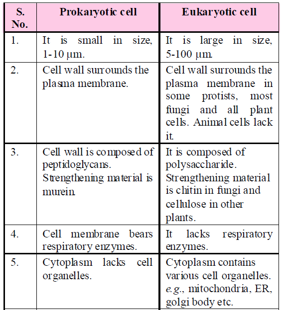
PROKARYOTIC CELLS
- They are typically unicellular organisms, e.g., bacteria, blue-green algae, mycoplasma and PPLO (Pleuropneumonia Like Organisms).
- The four basic shapes of bacteria are bacillus (rod like), coccus (spherical), vibrio (comma shaped) and spirillum (spiral).
- All prokaryotes have a cell wall surrounding the cell membrane. The fluid matrix filling the cell is the cytoplasm.
- The genetic material is naked circular DNA, not enclosed by a nuclear envelope.
- In addition to the genomic DNA (the single chromosome/circular DNA), many bacteria have small circular DNA called plasmids that confers certain unique phenotypic characters to such bacteria, e.g., resistance to antibiotics. Plasmid DNA is used to monitor bacterial transformation with foreign DNA.
- On the plasma membrane, there are some circular coiled bodies called mesosomes which are simply infoldings of the plasma membrane. They contain respiratory enzymes like oxidases and dehydrogenases and hence help in respiration. They help in cell wall formation, DNA replication and distribution to daughter cells.
CELL ENVELOPE AND ITS MODIFICATIONS
- The cell envelope consists of a tightly bound three layered structure i.e., the outermost glycocalyx followed by the cell wall and then the plasma membrane.
- Bacteria can be classified into two groups on the basis of the differences in the cell envelope and the manner in which they respond to the staining procedure developed by Gram viz., those that take up the gram stain are Gram positive bacteria (e.g., Streptococcus, Staphylococcus, Bacillus, Mycobacterium, Streptomyces etc.) and the others that do not are called Gram negative bacteria (e.g., Salmonella, Pseudomonas, Escherichia coli, Rhizobium, Helicobacter etc.)
- In a large number of bacteria, a thick slime capsule is present outside the cell wall which is made of polysaccharides and nitrogenous substances. It provides protection against phagocytosis and antibiotics.
- The plasma membrane is semi-permeable in nature and interacts with the outer surface. This membrane is structurally similar to that of the eukaryotes.
- In some prokaryotes like cyanobacteria, there are other membranous extensions into the cytoplasm called chromatophores which contain pigments like chla, β-carotene, myxoxanthophyll, myxoxanthin C- phycocyanin and C-phycoerythrin. In the presence of chlorophyll a, they synthesise their own food and certain others fix atmospheric nitrogen.
- Bacterial cells may be motile or non-motile. If motile, they have thin filamentous extensions from their cell wall called flagella. Bacterial flagellum is composed of three parts – filament, hook and basal body. The filament is the longest portion and extends from the cell surface to the outside.
- Besides flagella, pili and fimbriae are also surface structures of the bacteria but do not play a role in motility.
RIBOSOMES AND INCLUSION BODIES
- The cytoplasm is granular due to the presence of a large number of ribosomes (70s). Several ribosomes may attach to a single mRNA and form a chain called polyribosomes. They are the site of protein synthesis.
- Reserve materials are stored in the cytoplasm in the form of inclusion bodies, e.g., phosphate granules, cyanophycean granules and glycogen granules. Gas vacuoles are found in blue green and purple and green photosynthetic bacteria.
EUKARYOTIC CELLS
- The eukaryotes include all the protists (unicellular eukaryotes), plants, animals and fungi.
- An organised nucleus is present. It contains hereditary material covered on the outside by nuclear envelope.
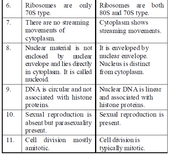
- All eukaryotic cells are not identical, e.g., plant and animal cells.
Differences between Plant and Animal cells
CELL MEMBRANE
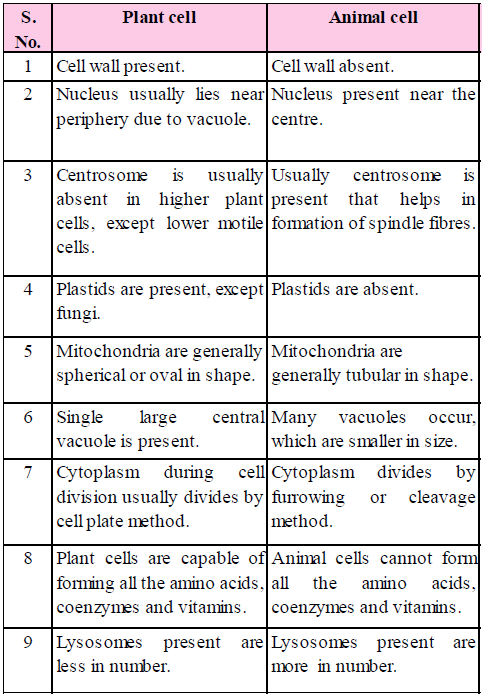
- Every living cell is externally covered by a thin, electron microscopic, elastic, regenerative and selectively permeable membrane called plasma membrane.
- Chemical composition : Lipoproteins, lipid and protein are the major components forming 60% of the plasma membrane. Proteins provide mechanical strength and are responsible for transportation of different substances. Proteins also act as enzymes. Lipids may account 28%-79% depending upon the type of cell and organism involved (in humans, myelin 79%). The lipids of plasma membrane are of three types namely phospholipids, glycolipids and sterols. The sterol found in the membrane may be cholesterol (animals), phytosterol (plants) or ergosterol (microorganisms).
- Carbohydrates form 2%-10%. The carbohydrates of plasma membrane are covalently linked to both lipid and protein components.
- An improved model of the structure of cell membrane was proposed by Singer and Nicolson (1972) widely accepted as fluid mosaic model. According to this, the quasi-fluid nature of lipid enables lateral movement of proteins within the overall bilayer.
- Fluidity of plasma membrane is due to phospholipids which is rich in unsaturated fatty acids. Phospholipids form the main component of cell membrane because it provides structural framework to the membrane.
- Lipids are amphipathic, i.e., they are structurally asymmetric with polar hydrophilic head and non-polar hydrophobic tail. On the outer side of some of the lipids, sugar chains are attached to their polar heads and hence are known as glycolipids.
- Two kinds of proteins are found in the membrane, i.e., the peripheral or extrinsic protein and the integral or intrinsic proteins.
- Many molecules can move briefly across the membrane without any requirement of energy and this is called the passive transport. Neutral solutes may move across the membrane by the process of simple diffusion along the concentration gradient, i.e., from higher concentration to the lower. Osmosis is the term used to refer specifically to the diffusion of water across a differentially or semi-permeable membrane.
As the polar molecules cannot pass through the non-polar lipid bilayer, they require a carrier protein of the membrane to facilitate their transport across the membrane. A few ions or molecules are transported across the membrane against their concentration gradient, i.e., from lower to higher concentration. Such a transport is an energy dependent process, in which ATP is utilised and is called active transport, e.g., Na+/ K+ Pump.
- Bulk transport : It is transport of large quantities of micromolecules, macromolecules and food particles through the membrane. It is accompanied by formation of transport or carrier vesicles.
- Pinocytosis : It is bulk intake of fluid, ions and molecules through development of small endocytotic vesicles of 100 – 200 nm in diameter. ATP, Ca2+, fibrillar protein (clathrin) and contractile protein (actin) are required.
- Phagocytosis : It is cell eating or ingestion of large particles by living cells, e.g., white blood corpuscles (neutrophils, monocytes), Kupffer’s cells of liver, reticular cells of spleen, histiocytes of connective tissues, macrophages, Amoeba and some other protists, feeding cells of sponges and coelenterates. Plasma membrane has receptors. As soon as the food particle comes in contact with the receptor site, the edges of the latter evaginate, form a vesicle which pinches off as phagosome.
- Functions
- It provides mechanical strength as well as acts as a protective layer.
- Plasma membrane is responsible for the transportation of materials, molecules, ions etc.
- Diffusion of gases (O2 and CO2) takes place through plasma membrane by simple and facilitated diffusion.
CELL WALL
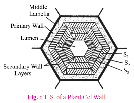
- Cell wall was discovered by Robert Hooke while observing the cell walls in cork tissue. It is the outermost, rigid, protective, non-living and supportive layer found in all plant cells, bacteria, cyanobacteria and some protists. It is not found in animal cells.
- A cell wall is organized at telophase stage of cell division. Fragments of ER and vesicles of golgi body align at the equator, termed as phragmoplast, later which forms the cell plate.
- Algae have cell wall made of cellulose, galactans, mannans and minerals like calcium carbonate, while in other plants it consists of cellulose, hemicellulose, pectins and proteins.
- In cell wall of a young plant cell, the primary wall is capable of growth, which gradually diminishes as the cell matures and the secondary wall is formed on the inner (towards membrane) side of the cell.
- Middle lamella is the outermost region which is found as a common cementing layer between two cells. It is formed of calcium and magnesium pectate.
- Pits are formed in lignified cell wall. They occur in sclerenchyma, vessels and tracheids. Tracheids in gymnosperms have maximum number of bordered pits.
- A number of plasmodesmata or cytoplasmic strands are present in pit through which the cytoplasm of one cell is in contact with another.
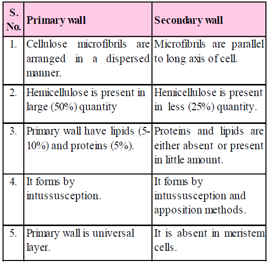
Differences between Primary wall and Secondary wall
- Growth of cell wall
- By intussusception : As the cell wall stretches in one or more directions, new cell wall material secreted by protoplasm gets embedded within the original wall.
- By apposition : In this method, new cell wall material secreted by protoplasm is deposited by definite thin plates one after the other.
- Function
- Cell wall not only gives shape to the cell and protects the cell from mechanical damage and infection, it also helps in cell-to-cell interaction and provides a barrier to undesirable macromolecules.
- Because of cell walls, plant cells can withstand a lot of variations in the surrounding medium as compared to animal cells.
CYTOPLASM
- The substance occur around the nucleus and inside the plasma membrane containing various organelles and inclusions is called cytoplasm.
- It forms about half of the cell’s volume and about 90% of it is water.
- It contains ions, biomolecules, such as sugar, amino acid, nucleotide, tRNA, enzymes, vitamins, etc.
- Cytoplasmic organelles are plastid, lysosome, sphaerosome, peroxisome, glyoxysomes, mitochondria, ribosome, centrosomes, flagella or cilia etc.
ENDOMEMBRANE SYSTEM
The endomembrane system includes endoplasmic reticulum (ER), golgi complex, lysosomes and vacuoles. Since the functions of the mitochondria, chloroplast and peroxisomes are not coordinated with the above components, these are not considered as part of the endomembrane system.
ENDOPLASMIC RETICULUM (ER)
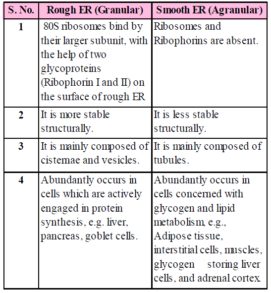
- A network or reticulum of tiny tubular structures scattered in the cytoplasm that is called the endoplasmic reticulum (ER).
- The ER is present in almost all eukaryotic cells. A few cells such as ova, embryonic cells, and mature RBCs, however, lack ER. It is also absent in prokaryotic cell. In rapidly dividing cells, endoplasmic reticulum is poorly developed.
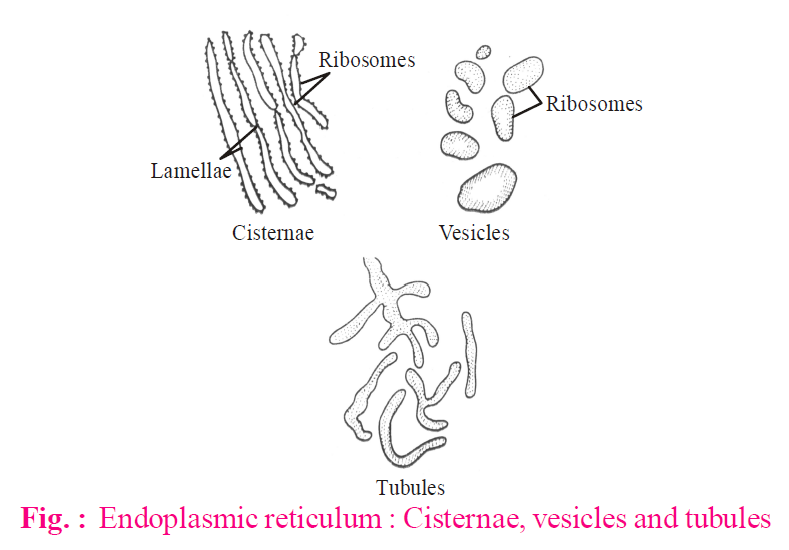
- The ER is made up of three components. All the three structures are bound by a single unit membrane.
- Cisternae : These are flattened, unbranched, sac like structures. They lie in stacks (piles) parallel to one another. They bear ribosomes.
- Vesicles : These are oval or rounded, vacuole like elements, scattered in cytoplasm. These are also studded with ribosomes.
- Tubules : Wider, tubular, branched elements mainly present near the cell membrane. They are free from ribosomes. These are more in lipid forming cells.
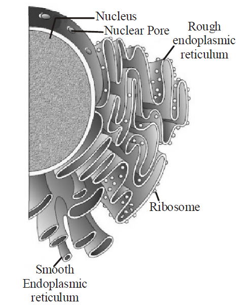
- The endoplasmic reticulum bearing ribosomes on their surface is called Rough Endoplasmic Reticulum (RER). In the absence of ribosomes, they appear smooth and are called Smooth Endoplasmic Reticulum (SER). RER is frequently observed in the cells actively involved in protein synthesis and secretion. The smooth endoplasmic reticulum is the major site for synthesis of lipid. In animal cells, lipid-like steroidal hormones are synthesised in SER.
Differences between Rough ER (Granular) and Smooth ER (Agranular)
- Functions
- Synthesis and secretion of specific proteins via-golgi bodies.
- Provides surface for synthesis of cholesterol, steroids, ascorbic acid, visual pigments and hormones e.g., testosterone and estrogen.
- ER helps in glycogenolysis in the liver cells and brings detoxification of many poisons and drugs (SER).
- ER is a component of cytoskeleton (spread as a net) of cell and provides mechanical support and shape to the cell.
GOLGI APPARATUS
- Camillo Golgi (1898) first observed densely stained reticular structures near the nucleus.
- These consist of many flat, disc-shaped sacs or cisternae of 0.5µm to 1µm in diameter. These are stacked parallel to each other.
- It is present in all eukaryotic cells. In plants, these are scattered irregularly in the cytoplasm and called as “dictyosomes”. These are absent in bacteria and blue green algae, RBCs, spermatozoa of bryophytes and pteridophytes, and sieve tube cells of phloem of angiosperm.
- Golgi body is made of 4 parts :
- Cisternae : Golgi apparatus is made up of stack or flat sac like structures called cisternae. The margins of each cisterna are gently curved so that the entire golgi body takes a cup like appearance. The golgi body has a definite polarity. The cisternae at the convex end of the dictyosome comprises forming face (F face) or cis face. While the cisternae at the concave end comprises the maturing face (M face) or trans face. The forming face is located next to either the nucleus or endoplasmic reticulum. The maturing face is usually directed towards the plasma membrane. It is the functional unit of golgi body.
- Tubules : These arise due to fenestration of cisternae and form a complex network.
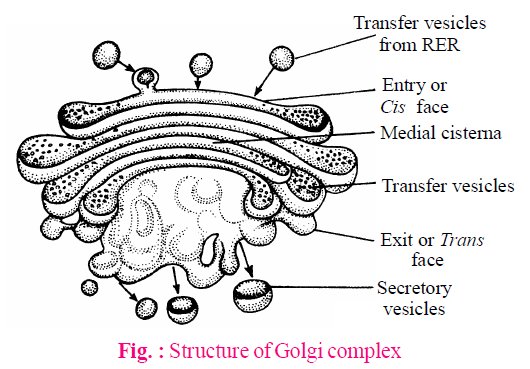
- Secretory vesicles : These are small sized components each about 40 Å in diameter present along convex surface of edges of cisternae. These are smooth and coated type of vesicles.
- Golgian vacuoles : They are expanded part of the cisternae which have become modified to form vacuoles.
- Functions
- The main function of golgi body is secretion, so it is large sized among the secretory cells.
- Glycosidation of lipids i.e., addition of oligosaccharides to produce glycolipids.
- Glycosylation of proteins i.e., addition of carbohydrate to produce glycoproteins.
- Helps in the formation of primary lysosomes.
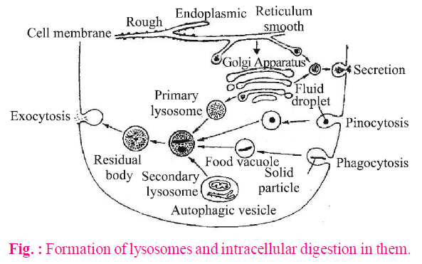
LYSOSOMES
- Lysosomes are microscopic, vesicular structures of the cytoplasm, bounded by a single membrane (lipoproteins) which are involved in intracellular digestive activities, containing hydrolytic enzymes. These are popularly called "suicidal bags".
- These were first discovered by a Belgian biochemist, Christian de Duve (1955) in the liver cells and named as "pericanalicular dense bodies".
- These are absent in prokaryotes but are present in all eukaryotic animal cells except mammalian RBCs. They have been recorded in fungi, Euglena, cotton and pea seeds.
- The isolated lysosomal vesicles have been found to be very rich in almost all types of hydrolytic enzymes (hydrolases – lipases, proteases, carbohydrases) optimally active at acidic pH. These enzymes are capable of digesting carbohydrates, proteins, lipids and nucleic acids.
- Types of lysosomes : On the basis of their contents, four types of lysosomes are recognised :
- Primary lysosomes : A newly formed lysosome contains enzymes only. Its enzymes are probably in an inactive state.
- Secondary lysosomes : When some material to be digested enters a primary lysosome, the later is named the secondary lysosome, or phagolysosome or digestive vacuole, or heterophagosome.
- Tertiary lysosomes/Residual bodies : A secondary lysosome containing indigestible matter is known as the residual bodies or tertiary lysosome.
- Autophagosomes/Autolysosomes : A cell may digest its own organelles, such as mitochondria, ER. This process is called autophagy. The acid hydrolases of lysosomes digest the organelles thus, it is called autophagosome.
- Functions
- Lysosomes of sperm provides enzyme for breaking limiting membrane of egg e.g., hyaluronidase enzyme.
- Lysosomes function as trigger of cell division or initiate cell division by digesting repressor molecules.
- They also engulf the carcinogens.
VACUOLES
- The vacuole in plants was discovered by Spallanzani. It is a non-living reservoir, bounded by a differentially or selectively permeable membrane, the tonoplast.
- These contain water, minerals and anthocyanin pigments.
- Some protozoans have contractile vacuoles which enlarge by accumulation of fluid or collapse by expelling them from the cell. The vacuoles may be sap vacuoles, contractile vacuoles or gas vacuoles (pseudo vacuoles).
- Functions
Vacuole maintains osmotic relation of cell which is helpful in absorption of water. Turgidity and flaccid stages of a cell are due to the concentrations of sap in the vacuole.
MITOCHONDRIA
- The mitochondria occur singly or in groups and their shape and size (0.5μ to 0.2μ) vary from cell to cell and depends on physiological activity of cell.
- Mitochondria is considered as semi-autonomous organelle because it has separate protein synthesizing machinery independent of nuclear control.
- These were first observed in striated muscles of insects as granules by Kolliker (1880), he called them "sarcosomes".
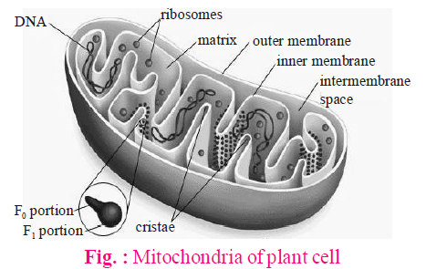
- Each mitochondrion is a double membrane-bound structure with the outer membrane and the inner membrane dividing its lumen distinctly into two aqueous compartments, i.e., the outer compartment and the inner compartment. The inner compartment is called the matrix. The outer membrane forms the continuous limiting boundary of the organelle. The inner membrane forms a number of infoldings called cristae towards the matrix. The cristae increase the surface area.
- Oxysomes have ATPase enzyme molecule and therefore, responsible for ATP synthesis. The reaction of ATP formation is endergonic. These elementary particles are also called F0 - F1 particles.
- The matrix also possesses single circular DNA molecule, a few RNA molecules, ribosomes (70S) and the components required for the synthesis of proteins. The mitochondria divide by fission.
- Functions
- Mitochondria are referred to as “powerhouse” of the cell as they produce 95% of ATP. This energy is produced during the breakdown of food molecules which involve glycolysis, oxidative decarboxylation and oxidative phosphorylation (krebs cycle and respiratory chain).
- Intermediate products of cell respiration are used in the formation of steroids, cytochromes, chlorophyll, etc.
- These are also seat of some amino acids biosynthesis.
- Mitochondria contain electron transport system.
PLASTIDS
- Plastids are semi-autonomous organelles having DNA, RNA, ribosomes and double membrane envelope. These are the largest cell organelles in plant cells.
- Haeckel (1865) discovered a plastid, but the term was first used by Schimper (1883).
- A well organised system of grana and stroma in plastid of normal barley plant was reported by de Von Wettstein.
- Park and Biggins (1964) gave the concept of quantasomes.
- Plastids are of 3 types: Leucoplasts, Chromoplasts and Chloroplasts.
LEUCOPLASTS
These are colourless plastids which generally occur near the nucleus in non-green cells and possess internal lamellae. These mainly store food materials and occur in the cells not exposed to sunlight, e.g., seeds, underground stems, roots, tubers, rhizomes etc.
These are of three types
- Amyloplast : Synthesize and store starch grains.
- Elaioplast (Lipidoplast, Oleoplast) : These store lipids and oils.
- Aleuroplast (Proteinoplast) : Store proteins.
CHROMOPLASTS
Coloured plastids other than green are known as chromoplasts. These plastids are red, orange, yellow etc. coloured due to the presence of carotenoid. These are present in petals and fruits.
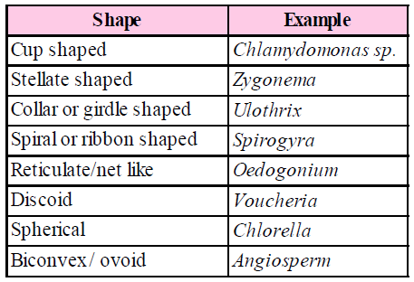
CHLOROPLAST
Discovered by Sachs and named by Schimper. Chloroplasts are green coloured plastids due to the presence of chlorophyll. These occur abundantly in green leaves and green parts of the shoot. These trap the solar energy which is used for manufacturing food. So, these are known as the sites of photosynthesis.
Shape : These have various shapes like
- It is a double membrane structure. Both membranes are smooth. The inner membrane is less permeable than outer but rich in proteins especially carrier proteins.
- The inter-membrane space is called the periplastidial space. Inner to membranes, matrix is present, which is divided into two parts–
- Grana : Inner plastidial membrane of the chloroplast is invaginated to form a series of parallel membranous sheets, called lamellae, which form a number of oval - shaped closed sacs called thylakoids.
Along the inner side of thylakoid membrane, there are a number of small rounded para-crystalline bodies called quantasomes (a quantasome is the photosynthetic unit).
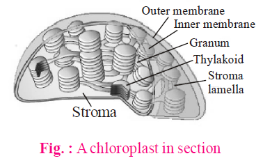
- Stroma : It is transparent, proteinaceous and watery substance. Dark reaction of photosynthesis occurs in this portion. Stroma is almost filled with "RuBisCO" (about 15% of total enzyme, protein) enzyme and CO2 is accepted by this enzyme. CO2 assimilation results in carbohydrate formation.
Chlorophyll a : C55 H72 O5 N4 Mg (with methyl group)
Chlorophyll b : C55 H70 O6 N4 Mg (with aldehyde group)
Chlorophyll c : C35 H32 O5 N4 Mg
Chlorophyll d : C54 H70 O6 N4 Mg
PIGMENTS OF CHLOROPLASTS
- Bacteriochlorophyll (C55H74O6Mg) or chlorobium chlorophyll present in photosynthetic bacteria.
- Carotenoids : These are hydrocarbons, soluble in organic solvents. These are of two types
- Carotenes : C40H56, derivatives of vitamin A. Carrot coloured α, β, γ carotene, lycopene, etc. β - carotene in the most common.
- Xanthophyll : C40H56O2, yellowish in colour, fucoxanthin, violaxanthin. Molar ratio of carotene and xanthophyll in young leaves is 2:1.
FUNCTIONS
- It is the site of photosynthesis (light and dark reactions).
- Photolysis of water, reduction of NADP to NADPH2 takes place in granum.
- Photophosphorylation through cytochrome b6 f, plastocyanin and plastoquinone etc.
- They store starch or factory of synthesis of sugars.
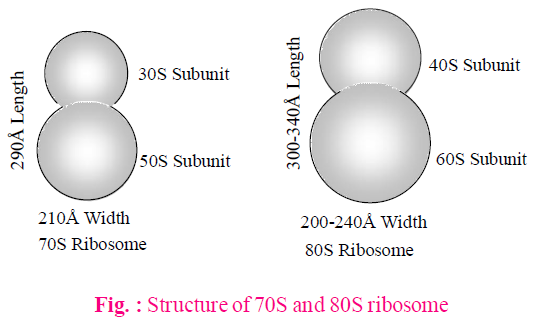
RIBOSOMES
The ribosomes are the smallest known organelles without membrane, ribonucleoprotein particles attached either on RER or floating freely in the cytoplasm and are the sites of protein synthesis.
TYPES OF RIBOSOMES
- 70S ribosomes : Found in prokaryotes, mitochondria and plastid of eukaryotes.
- 80S ribosomes : Found in cytoplasm of eukaryotes.
CHEMICAL COMPOSITION OF RIBOSOMES
70S (50S + 30S) (60S + 40S) - 60% r-RNA + 40% proteins
80S - 40% r-RNA + 60% proteins
60S - r-RNA 28S, 5.8S, 5S
40S - r-RNA 18S
50S - r-RNA 23S, 5S
30S - r-RNA 16S
Levine and Goodenough (1874) observed 77S ribosomes in fungal mitochondria, 60S ribosomes in animal mitochondria and 55S in mammalian mitochondria.
FUNCTIONS
- Ribosomes are also called protein factories of the cell.
- Enzyme peptidyl transferase occurs in large subunit of ribosome which helps in protein synthesis.
CYTOSKELETON
- In a eukaryotic cell, a framework of fibrous protein elements became necessary to support the extensive system of membranes.
- These elements collectively form cytoskeleton of the cell. These are of three types-
MICROTUBULES
These were first discovered by De Robertis and Franchi (1953) in the axons of medullated nerve fibres and were named neurotubules.
The microtubules are electron-microscopic structures found only in eukaryotic cellular structures like cilia, flagella, centrioles, basal-body, astral fibres, spindle fibres. These are mainly formed of tubulin protein.
FUNCTIONS
- These form a part of cytoskeleton and help in cell-shape and mechanical support.
- The microtubules of cilia and flagella help in locomotion and feeding.
- The microtubules of asters and spindle fibres of the mitotic apparatus help in the movement of chromosomes towards the opposite poles in cell-division.
MICROFILAMENT
• These were discovered by Paleviz et. al. (1974).
These are microscopic, long, narrow, cylindrical, non-contractile proteins found only in the eukaryotic cytoplasm. These are present in the microvilli, muscle fibres (called myofilaments) etc. But these are absent in prokaryotes. These are mainly formed of actin (contractile protein).
FUNCTIONS
- The microfilaments forms a part of cytoskeleton and change the cell shape, motility and division during development.
- The microfilaments bring about directed movements of particles and organelles along them in the cell.
INTERMEDIATE FILAMENTS
- These are supportive elements in the cytoplasm of eukaryotic cells. These are missing in mammalian RBCs.
- The IFs are somewhat larger than the microfilaments and are about 10 nm thick. These are solid, unbranched and composed of non-motile structural proteins, such as keratin, desmin, vimentin.
FUNCTIONS
- These form a part of cytoskeleton that supports the fluid cytosol and maintains the shape of the cell.
- These provide support to myofibrils which is essential for their contraction.
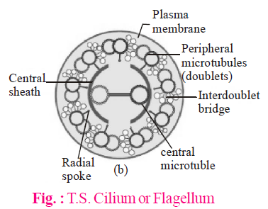
CILIA AND FLAGELLA
- Cilia (sing.: cilium) and flagella (sing.: flagellum) are hair-like outgrowths of the cell membrane. Cilia are small structures which work like oars, causing the movement of either the cell or the surrounding fluid. Flagella are comparatively longer and responsible for cell movement.
- Structure : Both cilia and flagella are structurally similar and possess similar parts - basal body, rootlets, basal plate and shaft.
- Basal body : It is present below the plasma membrane in cytoplasm. The structure is similar to centrioles made of 9 triplets of microtubules.
- Rootlets : Made of microfilament and provide support to the basal body.
- Basal plate : It is highly dense and lie above plasma-membrane.
- Shaft : It is the hair like projecting part of cilia and flagella which remains outside the cytoplasm. It has 9 doublet of microtubules in radial symmetry. These are called axonema. Each axonema has 11 fibrils, 9 in the periphery and 2 in the centre. The arrangement is called 9 + 2 pattern.
- Chemically, the central tubules are formed of dynein protein while the peripheral microtubules are formed of tubulin protein.
FUNCTIONS
- These help in locomotion, respiration, cleaning, circulation, feeding, etc.
- These show sensitivity to changes in light, temperature and contact.
Table : Differences between Cilia and Flagella

CENTROSOME AND CENTRIOLES
- Centrosome was first discovered by Van Benden (1887) and structure was given by T. Boweri.
- Centrosome is an organelle usually containing two cylindrical structures called centrioles. They are surrounded by amorphous pericentriolar materials. Both the centrioles in a centrosome lie perpendicular to each other in which each has an organisation like the cartwheel. They are made up of nine evenly spaced peripheral fibrils of tubulin. Each of the peripheral fibrils is a triplet.The adjacent triplets are also linked. The central part of the centrioles is also proteinaceous and called the hub, which is connected with tubules of the peripheral triplets by radial spokes made of protein.
- It has (9 + 0) pattern.
- Centriole is not covered by any membrane.
FUNCTIONS
- The centrioles help in organising the spindle fibres and astral rays during cell division.
- They provide basal bodies which give rise to cilia and flagella.
NUCLEUS
- It is the most important part of the cell which directs and controls all cellular functions.
- Nucleus as a cell organelle was first described by Robert Brown as early as 1831.
- Some mature cells even lack nucleus, e.g., erythrocytes of many mammals and sieve tube cells of vascular plants.
- A typical nucleus consists of the following structures :
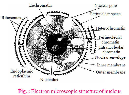
NUCLEAR MEMBRANE
- Nuclear membrane or nuclear envelope, consists of two parallel membranes with a space of about 10 to 50 nm called the perinuclear space which forms a barrier between the materials present inside the nucleus and that of the cytoplasm. The outer membrane usually remains continuous with the endoplasmic reticulum and also bears ribosomes on it.
- It is interrupted by minute pores which are formed by the fusion of its two membranes.
FUNCTIONS
- It allows the passage of inorganic ions, small organic molecules, ribosomal subunits, RNAs and proteins through nuclear pores.
- It maintains the shape of the nucleus.
NUCLEOLUS
- Nucleolus is a conspicuous, darkly stained spherical body found in the nucleoplasm.
- It is composed of large amount of ribosomal proteins and ribosomal RNA.
- It is generally associated with nucleolar organizer region (NOR) of the nucleolar chromosomes.
FUNCTIONS
- It is the seat of biogenesis of rRNA and also stores rRNA.
- It plays an important role in spindle formation during cell division.
NUCLEOPLASM
- It is a transparent, homogenous, semifluid, colloidal, ground substance present inside the nuclear membrane.
- It helps in maintaining the shape of nucleus, formation as well as spindle protein of NAD, ATP, DNA, RNAs and ribosomal subunits.
NUCLEAR MATRIX
It is a fine network of proteinaceous fibrils that traverses the whole nucleus.
FUNCTION
- It helps in maintaining shape of nucleus.
- It provides anchorage to chromatin.
CHROMATIN FIBRES / NUCLEAR CHROMATIN
These are thread like structures which are uniformly distributed in the nucleoplasm. These are observed only in the "interphase stage". Chromatin contains DNA and some basic proteins called histones, non-histone proteins and RNA.
FUNCTIONS OF CHROMATIN
- Chromatin stores genetic information.
- It forms chromosomes for equitable distribution of genetic information during cell division and reproduction.
- These are DNA-protein hereditary structures which are formed by condensation of chromatin fibres for equitable distribution during cell division and reproduction.
Every chromosome essentially has a primary constriction or the centromere on the sides of which disc shaped structures called kinetochores are present. Based on the position of the centromere, the chromosomes can be classified into four types :
- The metacentric chromosome has a centromere in the middle forming two equal arms of the chromosome.
- The sub-metacentric chromosome has centromere nearer to one end of the chromosome resulting into one shorter arm and one longer arm.
- In acrocentric chromosome, the centromere is situated close to its end forming one extremely short and one very long arm.
- The telocentric chromosome has a terminal centromere.
Sometimes a few chromosomes have non-staining secondary constrictions at a constant location. This gives the appearance of a small fragment called the satellite.
MICROBODIES
Many membrane bound minute vesicles called microbodies are present in both plant and animal cells that contain various enzymes.
SPHAEROSOMES
- These are found in all the plant cells which are involved in the synthesis and storage of lipids i.e., endosperm and cotyledon of oil seeds.
- These contain hydrolytic enzymes like protease, ribonuclease, phosphatase, esterase etc.
PEROXISOMES (URICOSOMES)
- These were called peroxisomes because these contain peroxide producing enzymes (oxidases) and peroxide destroying enzymes (catalases).
- These are found in photosynthetic cells of plants. In animals, peroxisomes are found in vertebrates (cells of the liver, kidney, brain, small intestine, testis and adrenal cortex), invertebrates and protozoans, e.g., Paramecium.
- Their membrane is permeable to amino acids, uric acids, etc. They contain four enzymes of H2O2 metabolism. The enzymes urate oxidase, d-amino oxidase, α-hydroxy acid oxidase produces H2O2 whereas the catalases play a significant protective role by degrading H2O2 because H2O2 is toxic for cells.
GLYOXYSOMES
- These are found in fungi, some protists and germinating fatty seeds where insoluble lipid food reserves must be turned into soluble sugars. These are absent in animal cells.
- These contain enzymes for the metabolism of glycolic acid via glyoxylate cycle and bounded by a unit membrane. These also contain enzymes for β-oxidation of fatty acids, producing acetyl CoA. It is metabolised in glyoxylate cycle to produce carbohydrates.
LOMASOMES
These are sac like structures found between cell wall and plasmalemma in the haustoria of fungal hyphae and spore producing structures, algal cells and in some cells of higher plants.
Study Notes for NEET/AIIMS/JIPMER


