


HUMAN REPRODUCTION
INTRODUCTION
- Reproduction is the process by which living organisms produces young one of their own type & reproductive system is a system of organs which takes part in this process.
- Humans are sexually reproducing and viviparous.
- Rate of reproduction is slower in sexual reproduction.
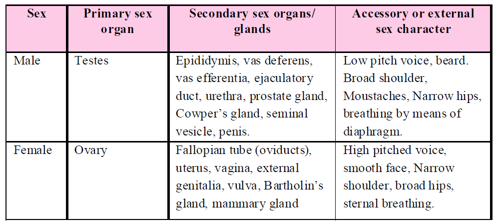
- Humans are unisexual. The reproductive system of each sex consists of many organs. The latter are distinguishable into primary and secondary sex organs. Besides these, there are some accessory sex characters.
- Primary sex organs : These are also called gonads which form gametes like - testis in males and ovary in females. Testis produces sperms and secretes testosterone. Ovary produces ova and secrete estrogen.
- Secondary sex organs : Sex organs, glands and ducts which do not produce gametes but are otherwise essential for sexual reproduction are known as secondary sex organs.
- Accessory / External / Secondary sex characters are traits which do not have any direct role in reproduction but provide specific features and structures to both the sexes.
- Beginning of sexual maturity or ability to reproduce is known as puberty. Puberty occurs at the age of 10 - 14 years in girls and 13 - 15 years in boys.
Table : Sexual characteristics in human beings
THE MALE REPRODUCTIVE SYSTEM
Male reproductive system is located in the pelvis region and consists of a pair of testis, a paired duct system consisting epididymis, vasa efferentia, vas deferens, ejaculatory duct & urethra.
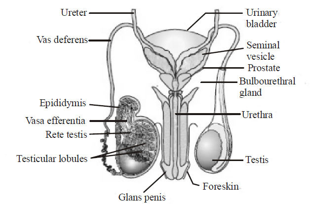
Fig. : Male reproductive system
TESTES
- The testes are situated outside the abdominal cavity within a pouch called scrotum.
- The scrotum allows sperms to develop at the optimum temperature (temperature of testes which is 2–2.5°C lower than the normal internal body temperature).
- The testis is attached to the scrotum by a band of connective tissue known as gubernaculum testis and the scrotum communicates with the abdominal cavity through inguinal canal.
- Each testis has about 250 compartments called testicular lobules. Each lobule contains one to three highly coiled seminiferous tubules in which sperms are produced.
- Each seminiferous tubule is lined by germinal epithelium which is formed of two types of cells -male germ cells (spermatogonia) and sertoli cells.
- Germ cells undergoes spermatogenesis to form spermatozoa and sertoli cell (also called subtentacular cells) functions as nurse cells for differentiating spermatozoa.
- The regions outside the seminiferous tubules (called interstitial spaces) contain small blood vessels and interstitial cells or Leydig cells. Leydig cells synthesize and secrete testicular hormones called androgens.
SECONDARY SEX ORGANS/GLANDS
- The seminiferous tubules of the testis open into the vasa efferentia through rete testis. The vasa efferentia leave the testis and open into epididymis located along the posterior surface of each testis.
- Epididymis is involved in temporary storage, nutrition & physiological maturation and motility of sperms. Epididymis is divided into three parts– anterior caput epididymis, middle corpus epididymis & porterior cauda epididymis (here spermatozoa is concentrated & stored until ejaculation).
- Ejaculation is the discharge of semen due to powerful rhythmic contraction of urethra.
- The duct system conduct the semen to the exterior.
- Vas deferens is a large duct which arises from the cauda epididymis & reach upto seminal vesicle.
- Ejaculatory ducts are short straight muscular tubes each formed by the union of vas deferens & duct of seminal vesicle. They have contractile mechanism that aids in the emission of seminal fluid.
- Urethra leads from the urinary bladder through the prostate glands and into the penis. Urethra has four parts– urinary, prostatic, membraneous and penile. The latter two form the outflow pathway for the urine and for the seminal fluid.
- Penis is the male copulatory organ. Glans penis is the tip of the penis which is highly sensitive to stimulation. Prepuce is a loose retractile foreskin which covers glans penis.
- Secretions of seminal vesicles, a prostate and paired bulbourethral glands constitute the seminal plasma which is rich in fructose, calcium and certain enzymes. The secretions of bulbourethral glands also helps in the lubrication of the penis.
- The seminal vesicles are long pouches with muscular wall. They secrete spermatozoa activating substances, such as fructose, citrate, inositol, prostaglandins and several proteins. Sperms use fructose as a respiratory substrate. Seminal fluid maintains viability and motility of sperms.
- Seminal vesicle secretes a alkaline, nutritive fluid which forms main part i.e., 60 % of the semen. It is also called uterus-masculinus. It forms the mullerian duct of the embryo.
NOTES
In females, these ducts form the oviducts. The seminal vesicle do not store sperms.
- Seminal vesicles are found in between the urinary bladder and rectum.
- The prostate gland surrounds the first portion of the urethra. This gland secretes a slightly acidic fluid (pH about 6.5) which forms 25% part of the semen. The secretion nourish and activates the spermatozoa to swim. It is essential for sperm motility (removal causes sterility).
- In the secretion of prostate gland, citric acid, calcium and phosphate, fibrinogen and fibrinolysin is present. The secretion of the prostate gland combines with the secretion of seminal vesicle and so the semen gets coagulated. In the coagulated semen, the mobility of sperms is reduced and so their energy is conserved. After sometime, due to fibrinolysins, semen again liquefies and in this semen, the sperms can move.
- Cowper's glands are also termed as Bulbourethral glands. 1st pair of Cowper's glands are attached to urethra. They secrete alkaline mucus which is discharged into the spongy part of urethra. The mucus lubricates the reproductive tract. This serves to neutralize any acid of urine remaining in the urethra. Secretion of Cowper's glands is produced before the ejaculation of semen.
- Secretion of Cowper's glands carries some spermatozoa released before ejaculation. This is one of the reasons for the high failure rate of the withdrawal method of birth control.
HORMONAL CONTROL OF MALE REPRODUCTIVE SYSTEM
The growth, maintenance and functions of secondary sex organs are under the control of testosterone hormone secreted by Leydig's cells of testis, while those of seminiferous tubules and Leydig's cells are controlled by Follicular Stimulating Hormone (FSH) and Interstitial Cells Stimulating Hormone (ICSH) of anterior pituitary lobe respectively.

Fig: Hormonal control of male reproductive system
THE FEMALE REPRODUCTIVE SYSTEM
The female reproductive system consists of a pair of ovaries, a pair of oviducts, a uterus, a vagina, external genitalia, and a pair of mammary glands.
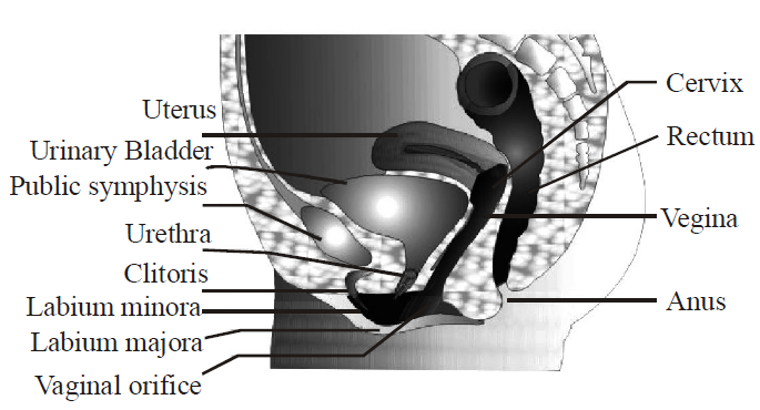
Fig. : Diagrammatic sectional view of female pelvis showing reproductive system
OVARY
- The ovaries produce the female gamete (ovum) and some steroid hormones (ovarian hormones).
- The ovaries are attached to the abdominal wall by an ovarian ligament called mesovarium.
- Each ovary is a compact or solid organ, consisting of an outer cortex and inner medulla. The stroma of the cortical region is composed of spindle shaped fibroblasts. A poorly delineated dense connective tissue layer, the tunica albuginea, covers the cortex. It imparts the whitish colour to the ovary. Located outside the tunica albuginea, the germinal epithelium formed of simple squamous or cuboidal epithelial cells covers the surface of the ovary.
Ovarian follicles in different stages of development are embedded in the stroma.
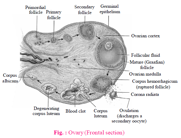
- Follicles are specialized structure in which oocyte growth & meiosis I occur.
- Mature follicles are known as Graafian follicle which occupy a single cavity called antrum & contains a secondary oocyte ready for ovulation.
- Ruptured graafian follicle is called corpus luteum (a temporary endocrine gland which secretes progesterone hormone for the maintenance of pregnancy).
- Corpus luteum loses its yellow colour & become inactive & transformed into a small cell mass called corpus albicans.
- Degenerated follicles are called atretic follicles.
NOTES
A female child at birth possess 80,000 follicles in the ovaries but about 400 mature & discharge their ova, the rest undergoes degeneration.
SECONDARY SEX ORGANS/GLANDS
- Oviducts develop from the mullerian duct of the embryo. It conveys the egg from the ovary to the uterus, and provides the appropriate environment for its fertilization. It is supported by a double fold of peritoneum called mesosalpinx.
- Each oviduct is differentiated into four parts – infundibulum (surrounded by finger like projections called fimbriae), ampulla (place where fertilization of ovum takes place), isthmus and uterine part.
- Fimbriae helps in collection of ovum after ovulation.
- Tubectomy is the cutting of oviduct & tying its two ends separately.
- Uterus (also called womb) is a pyriform, hollow muscular thick-walled but distensible median structure located above and behind the urinary bladder. It is meant for nourishing and development of foetus. For this, uterus is capable of tremendous enlargement. The empty uterus is 7.5 cm long and 5 cm broad and 2.5 cm thick.
- The surgical removal of uterus is called hysterectomy.
- The uterus has three layers – outer perimetrium, middle myometrium (thickest layer containing areolar connective tissue & smooth muscle fibres) & inner endometrium.
- During pregnancy, endometrium forms the maternal section of the placenta.
- The uterus is the site of implantation of the pre-embryo and for the subsequent embryonic & fetal development.
- The female external genitalia includes mons pubis, labia majora, labia minora, hymen and clitoris.
- Vagina is a tubular female copulatory organ and passageway for menstrual flow as well as birth canal. It is of about 10 cm length.
- Vaginal wall is made of an internal mucosa, muscular layer and an outer adventitia. Its mucous membrane is non-keratinized stratified squamous epithelium. Glands are absent. However, cervical glands do pass on some mucus into it during ovulation.
- During reproductive life, vagina contain certain bacteria (species of Lactobacillus and Lactoneustroc, also called Doderlein's Bacillus) which bring about fermentation and produce acid which inhibits the growth of other microorganisms.
In virgins, the vaginal orifice is partially covered by an annular centrally perforate membrane called hymen.
- Vulva ( = external female genitalia) is flanked by two pairs of fleshy folds of skin : the inner small, thin, moist, labia minora and outer larger, hair-covered labia majora ( = homologous to scrotal sac of male). All the labial folds have numerous sebaceous and sweat glands on both sides.
- A small erectile organ, the clitoris, lies at the anterior junction of the labia minora. It is homologous to the penis in the male but is very small and solid, having no passage through it. It consists of a short shaft with erectile tissue. It ends in a rounded glans clitori. The latter is covered by a small hook of skin, the prepuce. Rubbing of clitoris during intercourse produces a pleasurable sensation. This seems to be its only function.
- Urethra and vagina open by separate apertures, the upper urethral and lower vaginal orifices, into the vestibule. A fleshy elevation above the labia majora is known as mons veneris or mons pubis. It bears pubic hair, made up of adipose tissue.
- Bartholin's glands are a pair of small glands which open in the vestibule lateral to vaginal orifice. The secretion of this gland is thick, viscous and alkaline for lubrication and counteracting urinary acidity (similar to Cowper's glands in males).
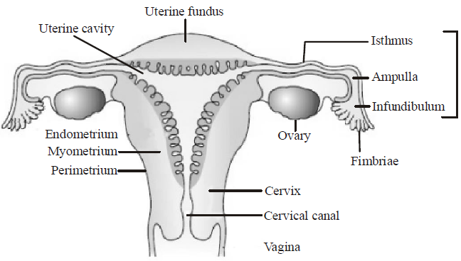
Fig. : Female reproductive system
- Mammary gland are modified sweat glands that lie over the pectoral muscle. Internally each gland contains 15-20 lobulated milk glands. Essential function of mammary gland is milk production which has nutritional and immunologic functions.
HORMONAL CONTROL OF FEMALE REPRODUCTIVE SYSTEM
- Ovary is regulated by pituitary gonadotropins or GnRH. Anterior pituitary secretes follicle stimulating hormone (FSH) which controls the transformation of young primary follicle into graafian follicle, maturation of ovum and secretion of estrogen by its follicular cells. The Luteinizing hormone (LH) of anterior pituitary regulates the ovulation from the graafian follicle, transformation of empty graafian follicle into yellowish, conical corpus luteum and secretion of progesterone hormone from the corpus luteum.
- Growth and function of secondary sex organs are regulated by estrogen and progesterone. Estrogen controls the growth, maintenance and functioning of secondary sex organs of female. Progesterone suspends ovulation during pregnancy, promotes implantation of foetus on the endometrium and development of foetus in the uterus.
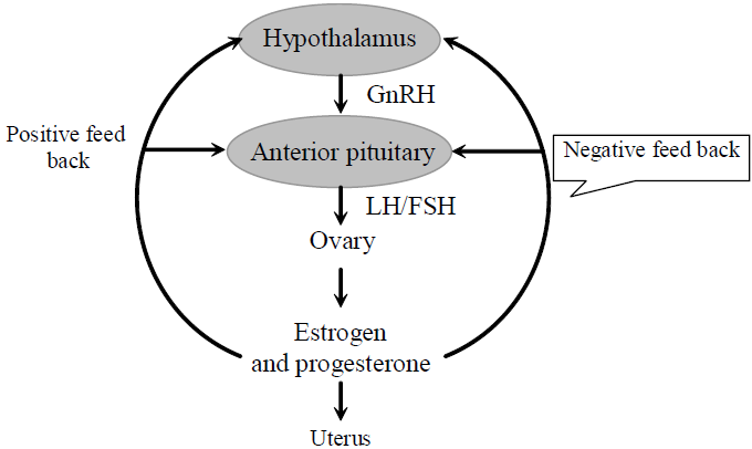
Fig. : Hormonal Control of Female Reproductive System.
- At the end of pregnancy, the corpus luteum secretes relaxin which broadens the pelvis for easy parturition.
GAMETOGENESIS
Gametogenesis is the process of gamete (sperm or egg) formation which include spermatogenesis & oogenesis.
SPERMATOGENESIS
- Spermatogenesis results in the formation of sperms that are transported by the male sex accessory ducts.
- Spermatogenesis is a continuous process & occurs in seminiferous tubules at the time of puberty (due to significant increase in the secretion of GnRH) & continues throughout life.
- Increased levels of GnRH acts at the anterior pituitary gland and stimulates the secretion of luteinizing hormone (LH) and follicle stimulating hormone (FSH). LH acts at the Leydig cells and stimulates synthesis and secretion of androgens. Androgens, in turn, stimulate the process of spermatogenesis. FSH acts on the Sertoli cells and stimulates secretion of some factors which help in the process of spermiogenesis.
- The spermatogonia present on the inner wall of seminiferous tubules multiply by mitotic division and increase in numbers. Each spermatogonium is diploid and contains 46 chromosomes. Some of the spermatogonia called primary spermatocytes periodically undergo meiosis.
A primary spermatocyte completes the first meiotic division (reduction division) leading to formation of two equal, haploid cells called secondary spermatocytes, which have only 23 chromosomes each.
The secondary spermatocytes undergo the second meiotic division to produce four equal, haploid spermatids.
- Spermiogenesis or spermateleosis is the process of formation of flagellated spermatozoa from spermatids.
- Spermiogenesis begins in the seminiferous tubules but usually completed in epididymis.
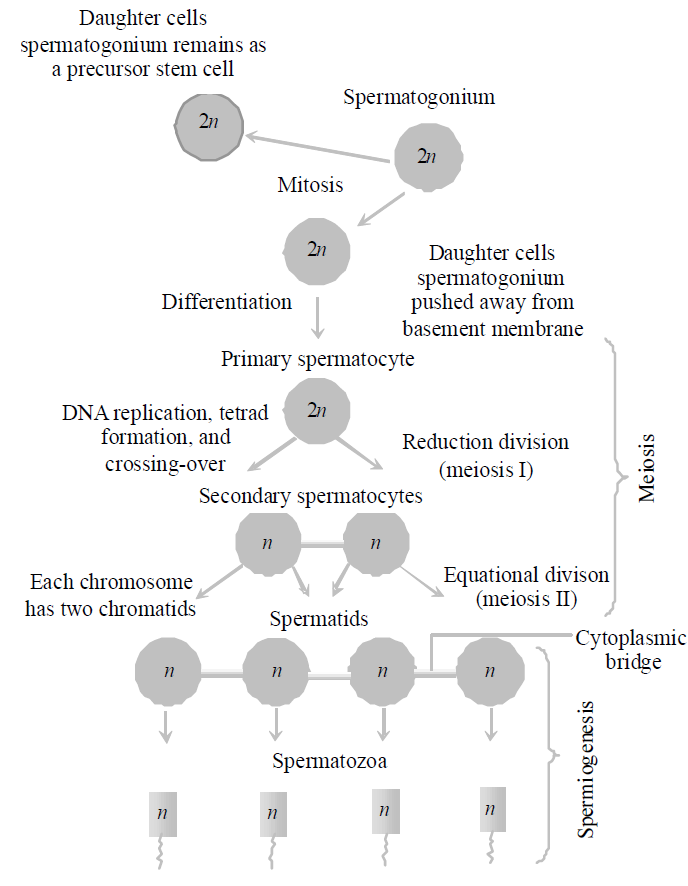
Fig. : Events in spermatogenesis
- After spermiogenesis, sperm heads become embedded in the sertoli cells, and are finally released from the seminiferous tubules by the process called spermiation.
STRUCTURE OF SPERM
- Sperm is a microscopic structure composed of a head, neck, a middle piece and a tail.
- The sperm head contains an elongated haploid nucleus, the anterior portion of which is covered by a cap-like structure, acrosome (produced by golgi body). The acrosome is filled with enzymes that help in fertilization of the ovum.
- If acrosome is removed from a sperm, it will fail to penetrate the ovum.
- Neck is very short containing proximal & distal centriole.
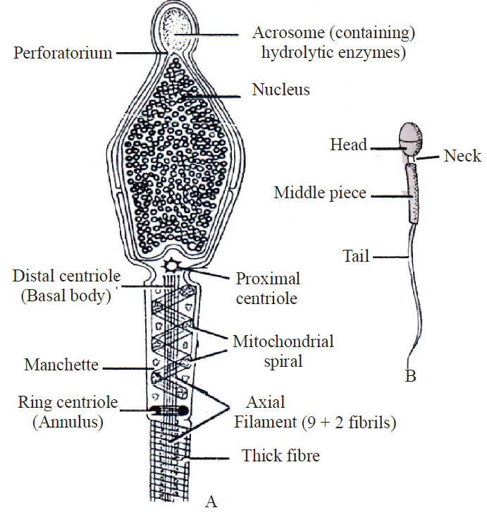
Fig. : Structure of sperm
- The middle piece possesses numerous mitochondria, which produce energy for the movement of tail that facilitate sperm motility essential for fertilization.
- Deficiency in the number of sperms result in sterility which is known as oligospermia.
- Absence of sperms in semen is known as azoospermia.
- Although normal number of sperm are present in semen but if these are completely non-motile, then this condition is known as necrospermia.
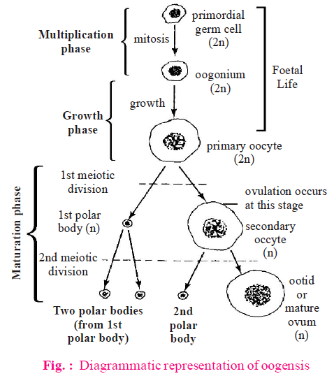
OOGENESIS
- The process of formation of a mature female gamete is called oogenesis.
- Oogenesis is a discontinuous process.
- Oogenesis begins before birth, stops in mid process and only resumes a year after menarche (the first menstrual bleeding).
- Oogenesis is initiated during the embryonic development stage when a couple of million gamete mother cells (oogonia) are formed within each fetal ovary; no more oogonia are formed and added after birth. These cells start division and enter into prophase-I of the meiotic division and get temporarily arrested at that stage, called primary oocytes.
- Each primary oocyte then gets surrounded by a layer of granulosa cells and then called the primary follicle. A large number of these follicles degenerate during the phase from birth to puberty. Therefore, at puberty only 60,000-80,000 primary follicles are left in each ovary.
- The primary follicles get surrounded by more layers of granulosa cells and a new theca and are called secondary follicles. The secondary follicle soon transforms into a tertiary follicle which is characterized by a fluid filled cavity called antrum.
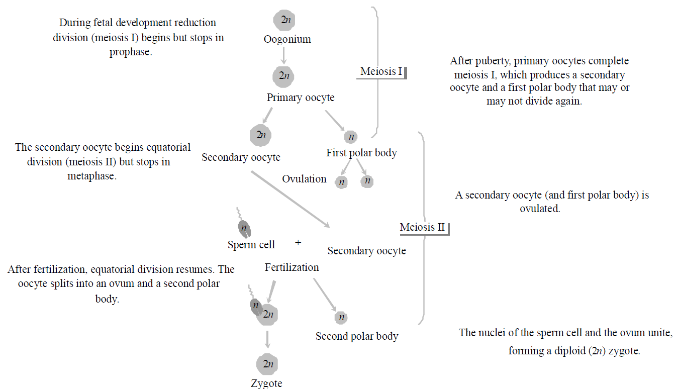
Fig. : Events in oogenesis
- The tertiary follicle further changes into the mature follicle or Graafian follicle. The secondary oocyte forms a new membrane called zona pellucida surrounding it. The Graafian follicle now ruptures to release the secondary oocyte (ovum) from the ovary by the process called ovulation.
NOTES
Secondary oocyte is a female gamete in which the 1st meiotic division is completed & second meiotic division (metaphase stage) begins. Secondary oocyte complete the secondary meiotic division only after fertilization by the sperm in the fallopian tube). The egg is released at secondary oocyte stage under the effect of LH.
- Oogenesis ends at menopause.
- Polar bodies are formed only in oogenesis at the time of formation of secondary oocyte.
STRUCTURE OF OVUM
- An ovum is generally spherical, non motile gamete with yolky cytoplasm and is enclosed in one or more egg.
Size of ovum varies in different animals and depends upon the amount of yolk. Size of ovum varies from 10µ to a few cm.
Egg size and yolk amount are interdependent. In mammals, it is generally microlecithal and about 100µ.
- The life span of eggs in female reproductive organs in human being is 48 hrs.
- The nucleus of egg is known as germinal vesicle.
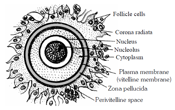
Fig. : Structure of Ovum
NOTES
Largest sized egg is of ostrich and is about 170 ×135 mm. Smallest egg is of humming bird.
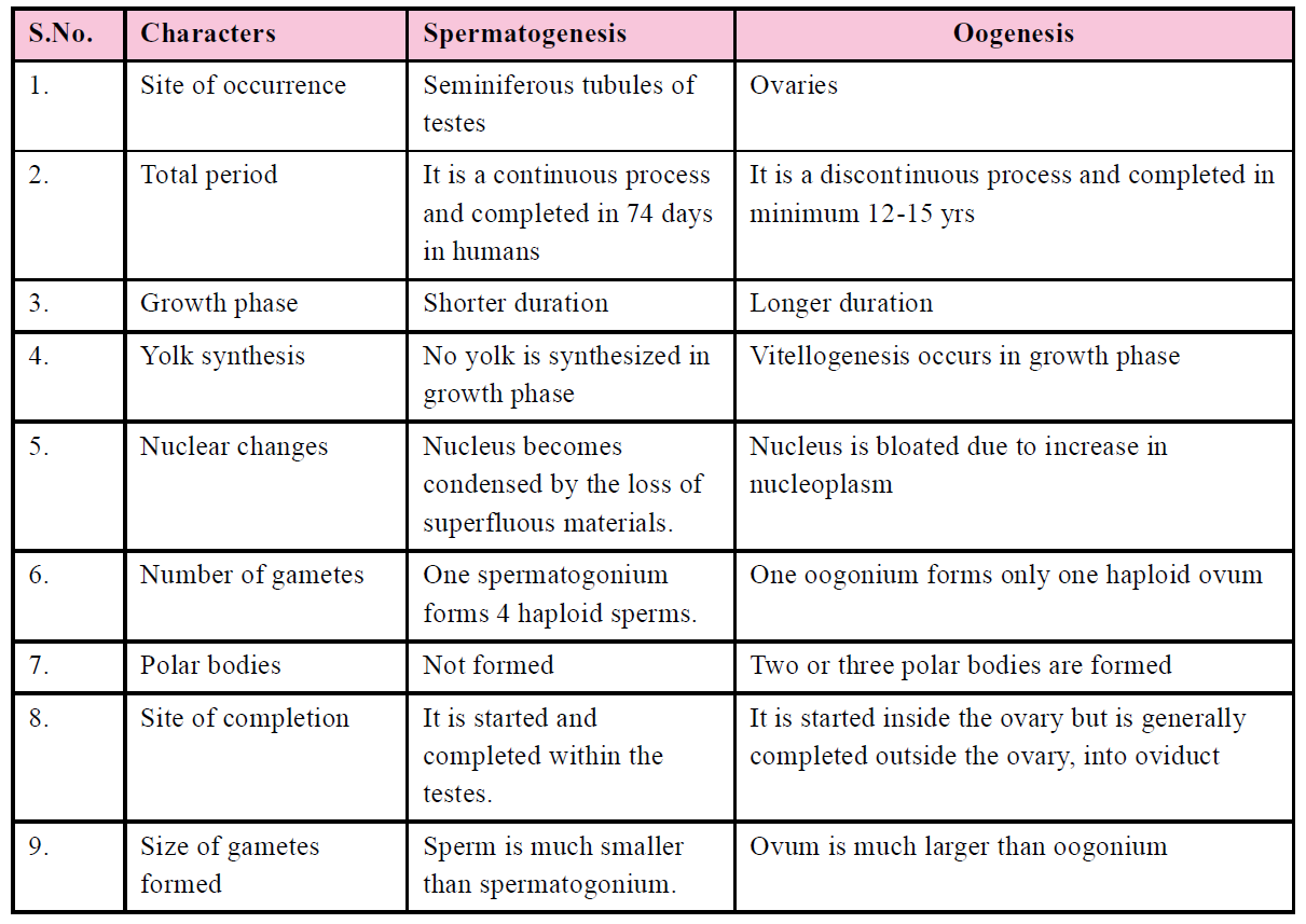
Table : Differences between Spermatogenesis and Oogenesis
MENSTRUAL CYCLE
- Menstrual cycle is the cyclic change in the reproductive tract of primate females.
- The first menstruation begins at puberty in females is known as menarche.
- Menstrual cycle starts only after attaining sexual maturation (puberty). During ovulation, only one ovum is released per menstrual cycle.
- The cyclical changes in the ovary and uterus during the menstrual cycle are induced by changes in the levels of pituitary and ovarian hormones.
- Cyclic menstruation is an indicator of normal reproductive phase and extends between menarche (the first menstrual cycle) and menopause.
- The length of menstrual cycle varies widely in women, but on average it is completed in 28 days (mensus means a month).
Menstrual cycle is absent during pregnancy, may be suppressed during lactation and permanently stops at menopause.
PHASES OF MENSTRUAL CYCLE
Menstrual cycle is divided into four phases - follicular, ovulatory, luteal and menstrual.
FOLLICULAR (PROLIFERATIVE) PHASE OR POST-MENSTRUAL OR PRE-OVULATORY PHASE
- It follows the menstrual phase and lasts for about 9-10 days (from 6 to 13th day of menstrual cycle).
- It involves following changes :
- Under the stimulation of FSH-RF of hypothalamus, there is increased secretion of FSH from anterior pituitary.
- FSH stimulates the change of a primary follicle of the ovary into a Graafian follicle.
- Follicular cells of Graafian follicle secrete estrogens.
- Proliferative phase consists of growth of endometrium, fallopian tube and vagina.
- The follicular phase ends with ovulation.
OVULATORY PHASE OR FERTILITY PHASE
- It involves the ovulation from the Graafian follicle of ovary.
- It occurs at the end of proliferative phase i.e. 14th day or midday during menstrual cycle and coincides with the beginning of the next phase.
- It is caused by increased turgidity and contraction of smooth muscles fibres around the Graafian follicle. Ovum is received by the fimbria of the fallopian tube. Ovum is viable for two days.
- Ovulation is controlled by the increased level of LH in the blood. Egg at that time is in the secondary oocyte state. LH also starts the change of empty Graafian follicle into corpus luteum and secretion of progesterone from corpus luteum.
- During ovulation, the secondary oocytes remains surrounded by its zona pellucida and corona radiata. There is no much change in uterine endometrium during ovulatory phase.
In animals, the ovulation follow three patterns :
- Fix or spontaneous ovulators : In these animals, ovulation takes place at a fix time in the midway of cycle. There is no need of coitus for ovulation. E.g., Primates (Human, Ape and Monkey).
- Induced or reflex ovulators : In these animals, copulation or coitus is necessary for ovulation, e.g., rabbit,
- Seasonal ovulators : Ovulation occur in breeding season, e.g., frog.
LUTEAL OR PROGESTATIONAL OR PRE-MENSTRUAL OR SECRETORY OR POST-OVULATORY PHASE
- It lasts for about 12 - 14 days and extends from 16th to 28th day of menstrual cycle.
- It is characterised by following changes -
- Corpus luteum (yellow body) is formed from empty Graafian follicle so is called luteal phase.
- The endometrium prepares for the implantation of an embryo & the corpus luteum is active.
- Corpus luteum begins to secrete a hormone called progesterone. The later reaches its peak about 22nd day after the beginning of cycle.
MENSTRUAL PHASE OR BLEEDING PHASE
- It lasts for about 3-5 days and extends from 1st to 4th day of the menstrual cycle.
- When the ovum remains unfertilized, then the corpus luteum starts degenerating. The level of progesterone in the blood declines. The uterine tissues fail to be maintained. Then the unfertilized ovum along with ruptured uterine epithelium, about 50 - 100 ml of blood and some mucus is discharged out through the vaginal orifice and is called menstrual flow or menstruation.
- Decrease in the level of progesterone and estrogen in the blood stimulates the hypothalamus and anterior pituitary to release FSH-RF and FSH respectively (positive feedback). FSH starts the follicular phase of next menstrual cycle.
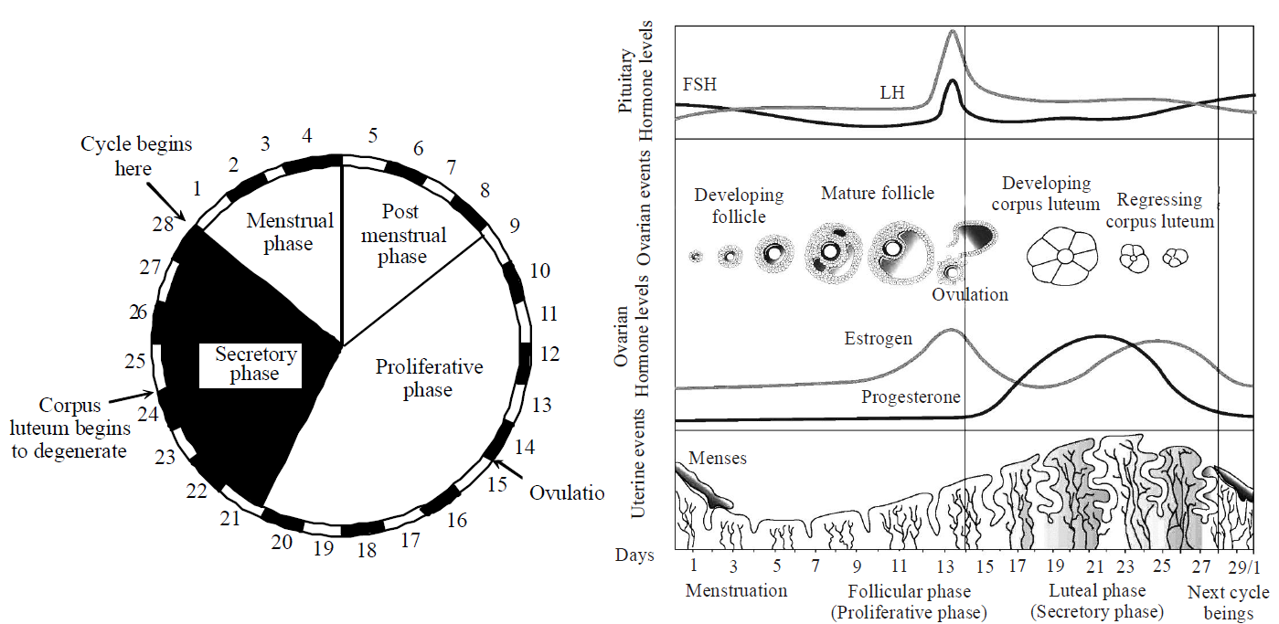
Fig. : Menstrual cycle and diagrammatic presentation of various events during a menstrual cycle
FERTILIZATION AND IMPLANTATION
- The process of fusion of a sperm with an ovum to form a diploid cell is called fertilization.
- Fertilization activates the secondary oocyte cell to complete the division.
- It normally occurs when sperm and egg interact in the upper part of the oviduct (ampulla)
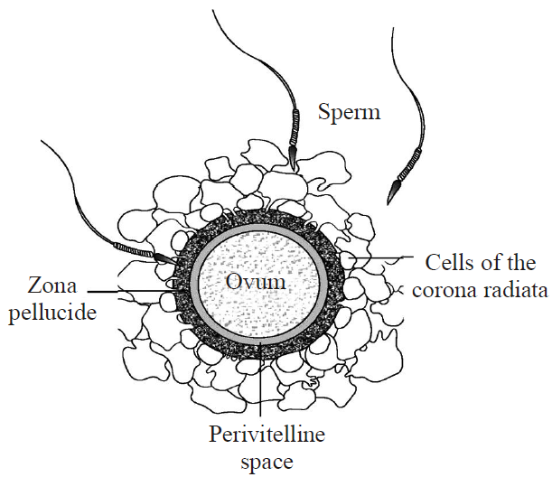
Fig. : Ovum surrounded by few sperms
- During fertilization, a sperm comes in contact with ovum (zona pellucida layer) and induces changes in the membrane that block the entry of additional sperms. Thus, it ensures that only one sperm can fertilize an ovum.
- Polyspermy is the entry of more than one sperm nucleus into an ovum at fertilization.
- When the acrosome of the spermatozoa touches the surface of egg, the cytoplasm of the egg bulges forward forming receptive cone or fertilization cone (a region where sperms enters the egg).
- The secretions of the acrosome help the sperm enter into the cytoplasm of the ovum through the zona pellucida and the plasma membrane. This induces the completion of the meiotic division of the secondary oocyte. The second meiotic division is also unequal and results in the formation of a second polar body and a haploid ovum (ootid). Soon the haploid nucleus of the sperms and that of the ovum fuse together to form a diploid zygote.
- Cleavage is the mitotic division of the zygote unit moving through the isthmus of the oviduct towards the uterus and forms 2, 4, 8, 16 daughter cells called blastomeres.
- Cleavage occurs more readily in the active cytoplasm.
- Cleavage in human is equal holoblastic.
- Morula is a solid ball of 32 cell stage without a cavity which is formed after 5th cleavage and 31 cell division. Morula looks like a little mulberry.
- Morula changes to blastula due to rearrangements of blastomeres.
- Blastula formation is called blastulation.
- Mammalian blastula with a large blastocoel is called blastocyst (in humans).
- Blastocyst has 3 parts-trophoblast, inner cell mass and blastocoel.
- The blastomeres in the blastocyst are arranged into an outer layer called trophoblast and an inner group of cells attached to trophoblast called the inner cell mass.
- The trophoblast layer then gets attached to the endometrium and the inner cell mass gets differentiated as the embryo.
- After attachment, the uterine cells divide rapidly and covers the blastocyst. As a result, the blastocyst becomes embedded in the endometrium of the uterus. This is called implantation (or nidation) and it leads to pregnancy.
- Implantation occurs generally between 6th to 9th day after fertilization.
- The site of implantation determines the portion of placenta.
- In human, implantation is of interstitial type in which embryo is buried in the uterine epithelium which completely surrounds it.
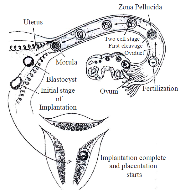
Fig. : Mechanism of Implantation
PREGNANCY AND EMBRYONIC DEVELOPMENT
- Placenta is an intimate mechanical connection between foetus and uterus of mother for nutrition, respiration and excretion.
- Placenta contains minute finger like projections called villi. The uterine wall forms corresponding depressions called crypts.
- Human placenta is hemochorial placenta (in which maternal blood bathes foetal chorionic villi directly), non-deciduous and meta discoidal.
- In simplest type of placenta, six barriers separate maternal blood from foetal blood. Three barriers are lost in human placenta.
- Umbilical cord is a flexible cord like structure connecting the foetus at the navel with the placenta and containing two umbilical arteries and one vein that nourishes the foetus and remove its wastes.
- Both type of blood vessels in the umbilical cord is surrounded by Whator's jelly.
- The placenta facilitates the supply of oxygen and nutrients to the embryo and also removal of carbon dioxide and excretory /waste materials produced by the embryo.
- Placenta also acts as an endocrine tissue as it secretes several hormones like human chorionic gonadotropin (hCG), human placental lactogen (hPL), estrogens, progestogens, etc that are essential for maintaining maternal physiological conditions appropriate for continued development of the conceptus. In the later phase of pregnancy, a hormone called relaxin is also secreted by the ovary.
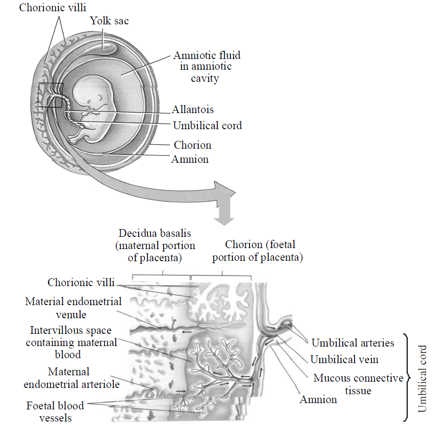
Fig : Placenta and Umbilical cord
- During pregnancy, the levels of other hormones like estrogens, progestogens, cortisol, prolactin, thyroxine, etc., are increased several folds in the maternal blood.
- Immediately after implantation, the inner cell mass (embryo) differentiates into an outer layer called ectoderm and an inner layer called endoderm. A mesoderm soon appears between the ectoderm and the endoderm. These three layers give rise to all tissues (organs) in adults.
- The establishment of germ layers initiates the final phase of embryonic development i.e. organogenesis.
DERIVATIVES OF ECTODERM
- Skin(epidermis) and their pigment cells.
- Mucosal membrane of lips, cheek, gums, basal portion of mouth, some part of palate, nasal apertures.
- Lower part of anal canal.
- Glans penis.
- Labia majora and outer part of labia minora.
- Anterior epithelium of cornea, epithelium of conjunctiva, ciliary body and iris of eyes.
- Outer face of tympanic membrane, epithelium of labyrinth.
- Glands-
- Exocrine : Sweat glands, sebaceous glands, parotid glands, mammary glands, lacrimal glands.
- Endocrine : Hypophysis cerebri and adrenal medulla.
- Hairs, nails, enamel of teeth
- Lens of eyes.
- Nervous system.
DERIVATIVES OF MESODERM
- Connective tissues, superficial and deep fascia, ligaments, tendons, dermis of skin (from dermatome)
- Specialized connective tissues like adipose tissue, reticular tissues, bones, cartilages.
- Teeth.
- All muscles.
- Heart, all blood vessels and blood cells.
- Kidneys, ureters, urinary bladder, posterior urethra of female, upper glandular part of prostate.
- Ovaries, uterine tubes.
- Testes, epididymis, vas deferens and seminal vesicle, ejaculatory duct.
- Pleural cavities, peritoneal cavity and pericardial cavity.
- Joints.
- Cornea, sclera, choroid ciliary body and iris related material.
- Microglia, dura mater etc.
DERIVATIVES OF ENDODERM
- Epithelial part of mouth, some part of palate, tongue, tonsils, pharynx, oesophagus, stomach, small and large intestine, upper part of anal canal.
- Pharyngo-tympanic tube, middle ear, inner face of tympanic membrane.
- Respiratory tract.
- Gall bladder, pancreatic duct.
- Major portion of urinary bladder, complete urethra of female except posterior part, complete urethra of male except anterior and posterior part.
- Whole inner part of vagina including inner face of labia minora.
- Glands-
- Exocrine : Liver and Pancreas
- Endocrine : Thyroid, parathyroid, thymus, islets of Langerhans.
- In addition to the above, the glands of gastrointestinal tract, major part of prostate etc. are also formed by endoderm.
PARTURITION AND LACTATION
- After nine months of pregnancy, the fully developed foetus is ready for delivery. The process of childbirth is called parturition.
- It is induced by a complex neuroendocrine mechanism involving cortisol, estrogens and oxytocin.
- Powerful contraction of the uterus in labor are needed for parturition.
- Stages of labor in parturition are
- 1st stage : Dilation of cervix
- 2nd stage : Delivery of baby
- 3nd stage : Delivery of placenta and umbilical cord.
- The signals for placenta parturition originate from the fully developed foetus and the placenta which induce mild uterine contraction called foetal ejection reflex.
- Foetal effection reflex induces oxytocin release from the posterior pituitary, which acts on uterine muscle and causes stronger uterine contraction.
- Gestation period is the length of time from conception to birth. Gestation period of 280 days is calculated from time of last menstruation (hence 266 days).
- Mammary glands differentiate during pregnancy and secrete milk after child-birth. The new-born baby is fed milk by the mother (lactation) during the initial few months of growth.
- The milk produced during the initial few days of lactation is called colostrum which contains several antibodies absolutely essential to develop resistance for the new-born babies.
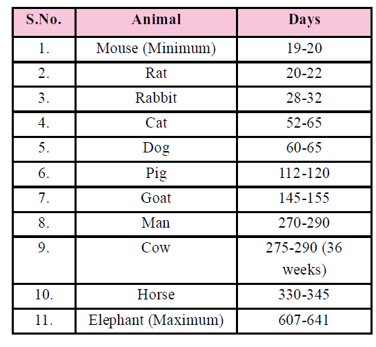
Table : Gestation period of different animals
Study Notes for NEET/AIIMS/JIPMER


