


BODY FLUIDS AND CIRCULATION
INTRODUCTION
- Body fluid is any fluid found within animals, including blood, lymph, tissue fluid, urine, bile etc.
- Circulation of body fluids are of two types–intracellular and extracellular circulation.
- Intracellular circulation occurs inside the cell through cyclosis, e.g., unicellular organisms like Paramecium, Amoeba, etc.
- Extracellular circulation occurs outside the body cell i.e. extracellular fluid that circulates in the body for transport of material.
- System which transport materials like nutrients, gases, hormones etc. to the various parts of the body and remove waste materials from the body cells is known as circulatory system.
- Study of circulatory system is called angiology.
- Father of angiology is William Harvey.
- The whole circulatory system is originated from mesoderm but the inner layer of the heart's wall (endocardium) and inner layer of blood vessel wall (endothelium) is endodermal in origin.
- Types of circulatory system are – open and closed circulatory system.
Table : Differences between open and closed circulatory system

BLOOD
- Blood is a fluid connective tissue that circulates through the heart, arteries, capillaries and veins carrying nutrients and oxygen to the body cells.
- Blood measures about 5-5.5 litres in an adult man constituting 30-35% of total extracellular fluid.
BLOOD COMPONENTS
Blood consists of plasma (fluid) and blood cells (formed elements).
PLASMA
- Plasma is a straw coloured, viscous fluid constituting nearly 55 per cent of the blood. 90-92 percent of plasma is water and proteins contributing 6-8 percent of it.
- Plasma contains three major classes of proteins - fibrinogen, globulins and albumins.
- Fibrinogens are needed for clotting or coagulation of blood. Globulins primarily are involved in defense mechanisms of the body and the albumins help in osmotic balance.
- Plasma contains small amounts of minerals like Na+, Ca++, Mg++, HCO3–, Cl–, etc. Glucose, amino acids, lipids, etc. are also present in the plasma as they are always in transit in the body. Factors for coagulation or clotting of blood are also present in the plasma in an inactive form.
- Plasma without the clotting factors is called serum.
- Plasma functions in
- transport
- body immunity
- prevention of blood loss
- retention of fluid in blood
- maintenance of blood pH
- uniform distribution of heat all over the body
BLOOD CELLS
- Blood cells are of three types : erythrocytes, leukocytes and platelets. These constitute nearly 45 per cent of the blood.
- Erythrocytes or red blood cells (RBC) are the most abundant of all the cells in blood.
- Process of RBC formation is called erythropoiesis (completed in 72 hours)
- A healthy adult man has, on an average, 5 millions to 5.5 millions of RBCs mm–3 of blood.
- RBCs are formed in the red bone marrow in the adults.
- RBCs are large microscopic cells without nuclei in mammals except camel and llama and are biconcave in shape.
- These have a red coloured, iron containing complex protein called haemoglobin, hence the colour and name of these cells.
- A healthy individual has 12-16 gms of haemoglobin in every 100 ml of blood. These molecules play a significant role in transport of respiratory gases.
- RBCs have an average life span of 120 days after which these are destroyed in the spleen (graveyard of RBCs).
- Function of RBC are-
- oxygen transport in the blood from the lungs to all the cells and tissues of the body.
- regulating the acid - base balance of blood, thus preventing large changes in pH.
- assist with the transport of carbon (IV) oxide from the tissues to the lungs.
- Leucocytes are also known as white blood cells (WBC) as these are colourless due to the lack of haemoglobin. WBC are nucleated and are relatively lesser in number which averages 6000-8000 mm3 of blood. Leukocytes are generally short lived.
- Two main categories of WBCs are - granulocytes and agranulocytes.
- Neutrophils, eosinophils and basophils are different types of granulocytes, while lymphocytes and monocytes are the agranulocytes.
- Neutrophils (bilobed nucleus) are the most abundant cells (60-65 per cent) of the total WBCs and basophils are the least (0.5-1 percent) among them.
- Neutrophils and monocytes (6-8 per cent) are phagocytic cells which destroy foreign organisms entering the body.
- Monocytes are the largest amongst all types of leucocyte with a rounded nucleus.
- Basophils secrete histamine, serotonin, heparin, etc., and are involved in inflammatory reactions.
- Eosinophils (2-3 per cent) resist infections and are also associated with allergic reactions.
- Lymphocytes (20-25 per cent) are of two major types - 'B' and 'T' forms. Both B and T lymphocytes are responsible for immune responses of the body.
- Platelets also called thrombocytes, are cell fragments produced from megakaryocytes (special cells in the bone marrow). Blood normally contains 1,500,00-3,500,00 platelets mm–3.
- Platelets can release a variety of substances most of which are involved in the coagulation or clotting of blood. A reduction in their number can lead to clotting disorders which will lead to excessive loss of blood from the body.
- Platelets have a lifespan of 9-10 days (about a week).
BLOOD GROUPS
- Blood groups are special characteristics of blood in humans and related primates due to the presence of genetically controlled antigens and antibodies.
- Two types of blood group are - ABO grouping & Rh grouping.
ABO GROUPING
- ABO grouping (was the first to be discovered) is based on the presence or absence of two surface antigens (chemicals that can induce an immune response) on the RBCs namely A and B. Similarly, the plasma of different individuals contain two natural antibodies (proteins produced in response to antigens).
- The distribution of antigens and antibodies in the four groups of blood, A, B, AB and O are :

- Group 'O' blood can be donated to persons with any other blood group and hence 'O' group individuals are called 'universal donors'.
- Persons with 'AB' group can accept blood from persons with AB as well as the other groups of blood. Therefore, such persons are called 'universal recipients'.
RH GROUPING
- There are several other proteins in the blood that may bring about agglutination under certain conditions. The most important of these is Rh factor.
- The Rhesus (Rh) system was discovered by Landsteiner and Weiner in 1940.
- Antigen, the Rh antigen similar to one present in Rhesus monkeys (hence Rh) is observed on the surface of RBCs of majority (nearly 80 per cent) of humans. Such individuals are called Rh positive (Rh+ve) and those in whom this antigen is absent are called Rh negative (Rh-ve).
- An Rh-ve person, if exposed to Rh+ve blood, will form specific antibodies against the Rh antigens. Therefore, Rh group should also be matched before transfusions.
- A special case of Rh incompatibility (mismatching) has been observed between the Rh-ve blood of a pregnant mother with Rh+ve blood of the foetus. Rh antigens of the foetus do not get exposed to the Rh-ve blood of the mother in the first pregnancy as the two bloods are well separated by the placenta. However, during the delivery of the first child, there is a possibility of exposure of the maternal blood to small amounts of the Rh+ve blood from the foetus. In such cases, the mother starts preparing antibodies against Rh in her blood. In case of her subsequent pregnancies, the Rh antibodies from the mother (Rh-ve) can leak into the blood of the foetus (Rh+ve) and destroy the foetal RBCs. This could be fatal to the foetus or could cause severe anaemia and jaundice to the baby. This condition is called erythroblastosis foetalis. This can be avoided by administering anti-Rh antibodies to the mother immediately after the delivery of the first child.
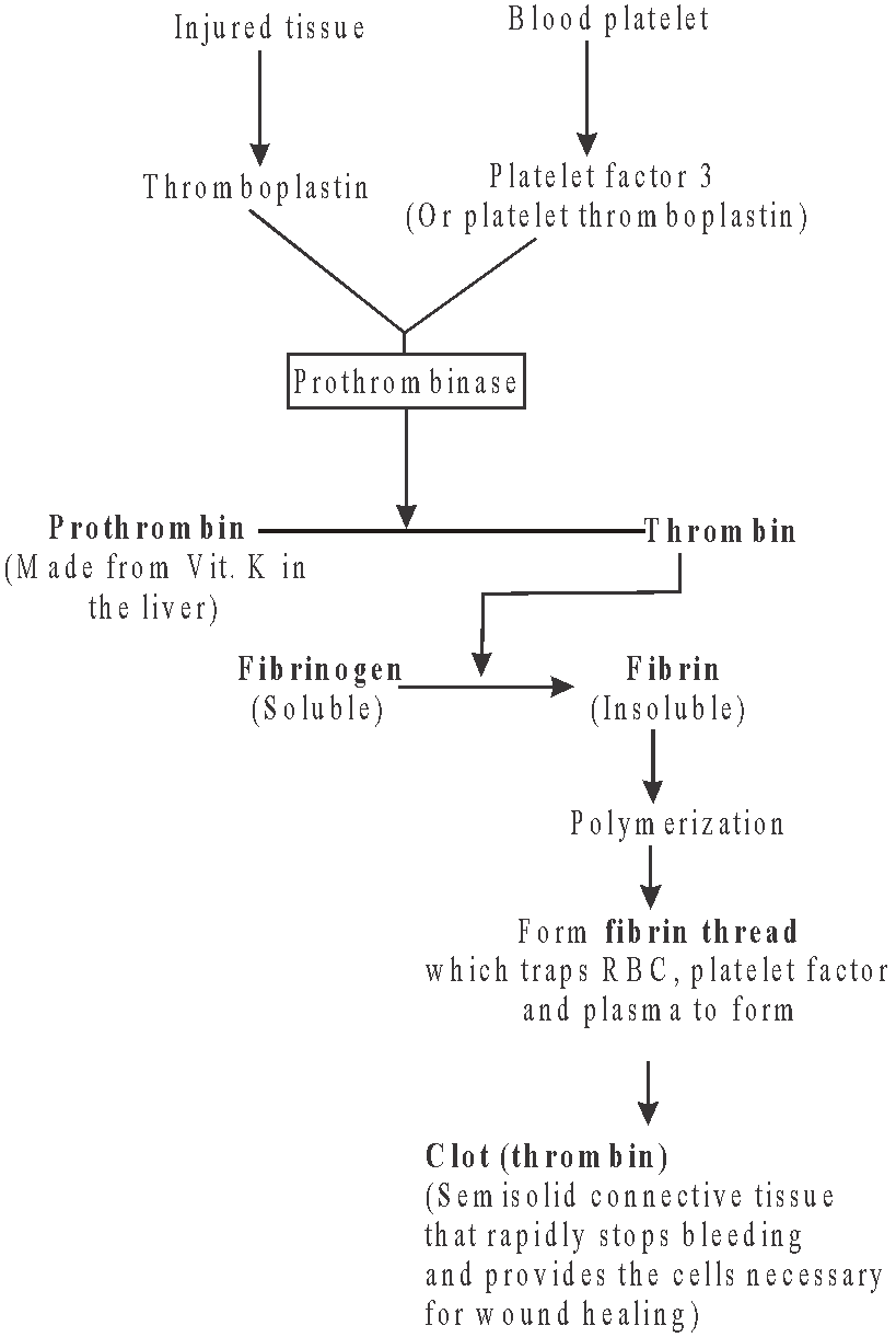
COAGULATION OF BLOOD
- It is the clotting of blood at the site of injury to prevent haemorrhage from damaged blood vessels.
- A clot formed mainly of a network of threads called fibrins in which dead and damaged formed elements of blood are trapped.
- Fibrins are formed by the conversion of inactive fibrinogens in the plasma by the enzyme thrombin. Thrombins, in turn are formed from another inactive substance present in the plasma called prothrombin. An enzyme complex, thrombokinase, is required for the above reaction.
- This complex is formed by a series of linked enzymic reactions (cascade process) involving a number of factors present in the plasma in an inactive state.
- An injury or trauma stimulates platelets in the blood to release certain factors which activate the mechanism of coagulation.
- Calcium ions play a very important role in clotting.
- The time taken for normal blood clotting varies from 4-10 minutes.
- Vitamin K is essential for normal clotting. It is given to a new born child immediately after the delivery to facilitate clotting of blood of the umbilical cord. A newborn baby has low levels of prothrombin & clotting factor due to low capacity to synthesize clotting factor compounded by deficiency of vitamin K.
Flow chart : Process of blood clotting
LYMPH
- Lymph is the interstitial fluid (tissue fluid present between the cells of a tissue.)
- Exchange of nutrients, gases, etc., between the blood and the cells always occur through this fluid.
- An elaborate network of lymph vessels (called the lymphatic system) collects this fluid and drains it back to the major veins.
- The fluid present in the lymphatic system is called lymph.
- Lymph is a colourless fluid containing specialised lymphocytes which are responsible for the immune responses of the body.
- Lymph is also an important carrier for nutrients, hormones, etc. Fats are absorbed through lymph in the lacteals present in the intestinal villi.
Lymph = Blood - [RBC + platelets + plasma proteins of high molecular weight]
- In humans, primary lymphatic (lymphoid) organs of the body are the red bone marrow and thymus gland. They are so called because they produce B and T cells, the lymphocytes that carry out immune response.
- Hematopoietic stem cells in red bone marrow gives rise to B cell and pre-T cells. Pre-T cells then migrate to the thymus gland.
- Secondary lymphatic organs are the lymph nodes and spleen.
LYMPH VESSEL
Structurally, these are similar to the veins, but in these thin wall and more valves are found than veins.
LYMPH NODES
- The lymphatic nodes (made up of lymphoid tissue) occur at intervals in the course of the lymphatic vessels.
- These contain lymphocytes, plasma cells and fixed macrophages.
- The lymph filters through the lymphatic nodes.
- The macrophages remove micro-organism, cellular debris and foreign particles from the lymph.
- Lymphatic nodes can detect and destroy cancer cells also.
- The lymphatic nodes also add lymphocytes and antibodies to the lymph.
SPLEEN
- The spleen is a large (7-10 cm. in diameter), bean-shaped, vascular, dark-red organ located in the abdomen just below the diaphragm at the tail of the pancreas behind the stomach.
- It is the largest lymph nodes and also called blood bank of body.
- It is the graveyard of RBC.
Table : Differences between lymph and blood

CIRCULATORY PATHWAYS
- The circulatory patterns are of two types - open or closed.
- All vertebrates possess a muscular chambered heart.
- Fishes have a 2-chambered heart with an atrium and a ventricle. The heart is called venous heart since it pumps deoxygenated blood to the gills for oxygenation. This blood goes directly from gills to visceral organs (single circuit circulation).
- Amphibians and reptiles (except crocodiles) have a 3-chambered heart with two atria and a single ventricle, whereas crocodiles, birds and mammals possess a 4-chambered heart with two atria and two ventricles.
- In amphibians and reptiles, the left atrium receives oxygenated blood from the gills/lungs/skin and the right atrium gets the deoxygenated blood from other body parts. However, they get mixed up in the single ventricle which pumps out mixed blood (incomplete double circulation).
- In birds and mammals, oxygenated and deoxygenated blood, received by the left and right atria respectively passes on to the ventricles of the same sides. The ventricles pump it out without any mixing up, i.e., two separate circulatory pathways are present in these organisms, hence, these animals have double circulation.
- Heart of aves consists of
- Left and right auricle
- Left and right ventricle
HUMAN CIRCULATORY SYSTEM
- Human circulatory system, also called the blood vascular system, consists of a muscular chambered heart, a network of closed branching blood vessels, blood and the fluid which is circulated.
- Circulatory system of human is of closed type.
HEART
- Heart is situated in the thoracic cavity between the lungs with its apex resting on the diaphragm.
- Usually, it is measured about 12 cm in length and 9 cm in breath.
- It is enclosed in double walled membranous bag, pericardium, enclosing the pericardial fluid.

HEART WALL
- The heart wall consists of connective tissue, blood vessels and cardiac muscle fibres in 3 different layers - epicardium, myocardium and endocardium.
- Epicardium : The outermost thin, transparent layer composed of mesothelium and connective tissue.
- Myocardium : It is the middle, highly vascular layer, composed of cardiac muscle fibres joined together by intercalated discs. The connective tissue in the myocardium acts as cardiac skeleton. Myocardium is thickest whereas the endocardium is thinnest.
Myocardium does not fatigue due to prolonged activity and formation of lactic acid.
- Endocardium : It is the innermost layer lining the cavity of the heart and consisting of endothelium of squamous cells resting on a thin basement membrane of loose connective tissue.
CHAMBERS OF HEART
- Heart has four chambers, with two anterior auricles and two posterior ventricles.
- The right atrium receives deoxygenated blood from the superior vena cava, inferior vena cava and coronary sinus. The left atrium receives oxygenated blood from two lungs through four pulmonary veins.
- A thin, muscular wall (called the interatrial septum) separates the right and left atria, whereas a thick-walled, (the interventricular septum) separates the left and the right ventricles.
- The walls of the ventricles are much thicker than that of the atria. Left ventricle is thicker than the right ventricle because it has to push blood to all body parts at a much greater pressure.
- The atrium and ventricle of the same side are also separated by a thick fibrous tissue called the atrio-ventricular septum. However, each of these septa are provided with an opening through which the two chambers of the same side are connected.
HEART VALVES
- The values of heart maintain unidirectional flow of blood and prevent its regurgitation in the opposite direction.
- Each valve has a set of cusps or flaps. The cusps are the folds of endocardium strengthened by an intervening layer of fibrous tissue.
- Opening and closing of valve depends upon pressure on opposite sides.
- When these valves break down, the blood does flow back and pool in the weakened legs resulting in varicose veins which often appear as large purplish tubes in the lower legs.
- The valves in the heart allows the flow of blood in only one direction, i.e., from the atria to the ventricles and from the ventricles to the pulmonary artery or aorta. These valves prevent any backward flow.
- Types of valves are - tricuspid, bicuspid, semilunar valve, etc.
- The opening between the right atrium and the right ventricle is guarded by a valve formed of three muscular flaps or cusps, the tricuspid valve, whereas a bicuspid or mitral valve guards the opening between the left atrium and the left ventricle.
- The bicuspid valve is also called as Mitral valve .
- Strong fibrous strings connecting bicuspid and tricuspid valves are known as chordae tendineae.
- The openings of the right and the left ventricles into the pulmonary artery and the aorta respectively are provided with the semilunar valves. These allow the passage of blood from the ventricles to respective blood vessels, but prevent the return of blood.
- Eustachian valve is present on the opening of inferior vena cava (post caval) in the right auricle in rabbit, whereas in human, the vestige of eustachian valve is present over the opening of post caval vein. It allows the passage of blood in right auricle.
- Haversian valve is present in human but absent in rabbit. It is present over the opening of precaval vein and allows the passage of blood in right auricle.
- Thebesian or coronary valve is present over the opening of coronary sinus in right auricle in mammals and allows the passage of blood in right auricle.
AUTOMATIC RHYTHMICITY OF HEART
- Automatic rhythmicity of the heart is the ability to contract spontaneously and at a regular interval of time.
- A specialised cardiac musculature called the nodal tissue is also distributed in the heart. A patch of this tissue is present in the right upper corner of the right atrium called the sino-atrial node (SAN). Another mass of this tissue is seen in the lower left corner of the right atrium close to the atrio-ventricular septum called the atrio-ventricular node (AVN).
- A bundle of nodal fibres, atrioventricular bundle (AV bundle) continues from the AVN which passes through the atrio-ventricular septa to emerge on the top of the interventricular septum and immediately divides it into a right and left bundle.
- Purkinje branches give rise to minute fibres throughout the ventricular musculature of the respective sides and are called purkinje fibres. Purkinje fibres along with right and left bundles are known as bundle of HIS.
- The nodal musculature has the ability to generate action potentials without any external stimuli, i.e., it is auto-excitable. However, the number of action potentials that could be generated in a minute vary at different parts of the nodal system.
- The SAN can generate the maximum number of action potentials, i.e., 70-75 min–1, and is responsible for initiating and maintaining the rhythmic contractile activity of the heart. Therefore, it is called the pacemaker.
HEART BEAT
- Heart beat is the rhythmic contraction and relaxation of the heart which include one systole (contraction phase) and one diastole (relaxation phase).
- Heart normally beats 70-75 times per minute (average 72 beats min–1) in normal adult human.
- Less number of heart beat than normal is called bradycardia.
- More rate of heart beat than normal is called tachycardia.
- Rate of heart beat increases :
- After taking food
- During exercise
- Decrease in blood pH
- Increased acidity and CO2 concentration
- Increase in temperature
- Tension/shock
- In high B.P.
- Regulation of heart beat : The centre controlling the heartbeat (cardiac centre) is present in medulla oblongata of brain and possess chemoreceptors sensitive for CO2, O2 and also for blood pressure. This centre is under the influence of hypothalamus which controls autonomic activities.
- In nervous control, brain receives two sets of nerve fibres- sympathetic and parasympathetic or vagal.
- When there is increase in blood CO2, the sympathetic nerve fibres stimulate S.A. node by producing sympathin (adrenaline + noradrenaline). This compound induces impulse generation by inducing entry of Ca2+ into cardiac muscles. So, heart beat and force of contraction increases (tachycardia). After action, sympathin is destroyed by sympathenase, COMT (catechol orthomethyl transferase) and MAO (Mono Amino Oxidase).
- When there is an increase in blood O2, the parasympathetic or vagus (10th cranial) nerve inhibits S.A. node by producing acetylcholine. This compound increases contraction time and hence, heart beat is decreased (bradycardia). After action, acetylcholine is destroyed by enzyme acetylcholinesterase (AchE).
- Stimulation of vagus nerve decreases the heart rate but its continuous stimulation shows no further decrease. This phenomenon is called vagus escape.
- Hormonal control : Hormones from adrenal medulla (adrenaline and noradrenaline) accelerate the heart beat, the latter under normal conditions and the former at the time of emergency. Thyroxine hormone also increases the heart beat by increasing energy production.
- Pounding refers to very fast heart beat during some conditions like anger and love.
- Increase in Na+ ions in blood or in cardiac muscle, decrease heart rate.
- Increase in Ca2+ ions in blood increase heart beat but if they are injected in cardiac muscles, heart stops in contracted phase which is called systolic arrest.
- Injection of K+ ions in heart muscles stop impulse generation. So, heart stops in diastolic or relax phase.
- On the basis of origin of heart beat, two types of heart are - neurogenic & myogenic
- In neurogenic heart, beat is initiated by a nerve impulse coming from a nerve ganglion situated near the heart. So in this, wave of contraction is generated outside the heart in the ganglion. If nerve supply is cut off then heart beat stops. E.g., Invertebrates (some annelids, most arthropods)
- In myogenic heart, beat is originated by a group of muscle fibres which is situated in the wall of the heart. So, in this wave of contraction is generated inside the heart. E.g., vertebrates, mollusca.
BLOOD VESSELS
- Blood vessels are intricate network of tubes that transport blood throughout the body. Blood vessels are made up of three layers – tunica externa (outermost), tunica media (middle) and tunica interna (innermost).
- The blood vessels are of following types: arteries, veins and capillaries.
ARTERIES
- Arteries are thick walled, carrying oxygenated blood (deoxygenated in pulmonary artery) from the heart to various parts of the body.
- These blood vessels are grouped as aorta which branches to form arteries which further divides into thinner branches called arterioles inside the organ.
- Average diameter of arteriole is 120µm. The arterioles further divide into smaller vessels called meta-arterioles (70 µm) which divide into capillaries. At the beginning of capillary, the arterioles possess circular muscles called pre-capillary sphincter which regulates flow of blood into the capillaries which is called vasomotion.
- Smooth muscles of arteries innervated by sympathetic fibers, their stimulation control vasoconstriction and vasodilation.
- Smooth muscles of arteries and arterioles also limit bleeding from wounds by producing vascular spasm during cut.
- Arteries are of two types–
- Conducting or elastic arteries
- Distributing or muscular arteries
- Elastic or conducting arteries receive blood from the heart and do not provide it to any organ rather these provide blood to other arteries and are pressure reservoirs of blood.
- Muscleless end of meta-arteriole is called thoroughfare channel or preferential channel.
- The largest artery is dorsal / abdominal aorta (systemic aorta).
CAPILLARIES
- These are the smallest blood vessels, discovered by Marcello Malpighi (also layered with nucleated squamous epithelial cells called endothelium resting on a basement membrane).
- Diameter of capillary is about 8nm .
- These are also called as exchange vessels as these are the site of exchange of material between blood and tissue because of least barrier in them.
- The capillaries can be grouped into two categories:
- arteriolar capillaries, which supplies nutrition, respiratory gases etc. to the body cells.
- veinular capillaries, which collect the metabolic wastes from the body cells.
- Capillaries possess about 7% of total body blood and are present in almost all cells of body in the intercellular spaces. The tissues which are devoid of intercellular spaces are also devoid of capillary. These are called avascular tissues.
VEINS
- These are thin walled, carrying deoxygenated blood (oxygenated in pulmonary vein) from tissues to the heart.
- Venules, smallest branches, unite to form veins which in turn unite to form vena cava.
- The largest vein is inferior vena cava/post caval.
- Inferior vena cava or post caval drains deoxygenated blood from middle and lower parts of the body into the right auricle through a single opening which is bordered by a membranous, falciform fold which is a remnant of the foetal valve of Eustachian.
- Superior vena cava or precaval : Brings deoxygenated blood from head and upper parts of the body into the right auricle through an opening which is single in human and cat and two in rabbit as there are 2 precavals - right and left in rabbit.
Table : Differences between Arteries and veins

CARDIAC CYCLE
- Cardiac cycle refers to the sequence of events which takes place during the completion of one heartbeat.
- The action potential causes the atria and then the ventricles to undergo contraction (systole) followed by their relaxation (diastole). The systole forces the blood to move from the atria to the ventricles and to the pulmonary artery and the aorta.
- In human being, the cardiac cycle occurs about 72 times per minute.
- Cardiac cycle is completed in following phases -
- Isometric relaxation
- Atrial diastole
- Ventricular systole
- Ventricular diastole
- Cardiac cycle is completed in 0.88 sec.
- About 70 mL of blood is pumped out by each ventricle during the cardiac cycle and it is called the stroke or beat volume.
- Volume of blood pumped out by each ventricle of heart per minute is called the cardiac output and it is equal to the product of stroke volume and heart rate (approx 5 litres).
- During each cardiac cycle, two prominent sounds are produced which can be easily heard through a stethoscope. The first heart sound (lubb) is associated with the closure of the tricuspid and bicuspid valves whereas the second heart sound (dupp) is associated with the closure of the semilunar valves.
- The first sound has a duration of 0.15 second and a frequency of 25 - 45 Hz. The second sound lasts about 0.12 seconds with a frequency of 50 Hz.
- Murmurs are abnormal sounds heard in various parts of vascular system. It may arise due to improper closing of any heart valve or in patients with inter-ventricular septal defects.
BLOOD PRESSURE
- The pressure exerted by the blood on the wall of the blood vessels in which it is present is called blood pressure.
- It is usually measured in the brachial artery by an instrument called a sphygmomanometer.
- Arterial blood pressure is of 2 types : Systolic and Diastolic.
- Systolic blood pressure is the pressure exerted by blood on the walls of the blood vessels due to the systole of the ventricles and is equal to 120 mm Hg. During ventricular systole, there is expansion in the artery due to the uncoiling of elastic layer. Hence, the pressure is maximum in arteries but gradually decreases in capillaries and veins.
- Diastolic blood pressure is the pressure exerted on the walls of blood vessels when the ventricles are relaxed. During ventricular diastole, the uncoiled elastic layer recoils leading to normalization of artery. Hence, blood pressure drops down to 80 mm Hg.
- Thus, blood pressure in normal person is systolic/diastolic pressure i.e. 120/80 mm Hg.
- The difference between systolic and diastolic pressures is called pulse pressure and its normal value is 120 - 80 mm Hg = 40 mm Hg. It provides information about the condition of arteries.
ELECTROCARDIOGRAM (ECG)
ECG is a graphical representation of the electrical changes that accompany cardiac cycle. These changes can be recorded with the help of an apparatus called electrocardiograph.

Fig. : Standard ECG
- Normal ECG is composed of P-wave, QRS wave (complex) and T-wave.
- The P-wave (a small upward wave) represents the electrical excitation (or depolarisation) of the atria, which leads to the contraction of both atria. It is caused by activation of SA node.
- QRS wave begins as a small downward deflection (R) and continues as a large upright (R) and triangular wave ending as downward wave (S) at its base. The QRS complex represents the depolarisation of the ventricles, which initiates the ventricular contraction. The contraction starts shortly after Q and marks the beginning of the systole.
- The T-wave represents the return of the ventricles from excited to normal state (repolarisation). The end of the T-wave marks the end of systole.
- Heart beat rate of an individual can be determined by counting the number of QRS complexes that occur in a given time period.
- Since the ECGs obtained from different individuals have roughly the same shape for a given lead configuration, any deviation from this shape indicates a possible abnormality or disease.
- Enlargement in Q & R wave indicates myocardial infarction.
- T-wave is flat when the heart muscles receive insufficient oxygen as in other sclerotic heart disease. It may be elevated, when the body's potassium level is increased.
DOUBLE CIRCULATION
- Double circulation is the passage of blood twice in the heart through separate pathways for completing one cycle.
- It is present in lung fishes, amphibians, birds, reptiles and man where atriovenous heart (means when it receives both venous and deoxygenated and arterial or oxygenated blood) is present.
- Double circulation consists of pulmonary and systemic circulation.

- In pulmonary circulation, right portion of heart collects impure blood from the body and sends it into the lungs. So in the right portion, impure blood is present and this circulation takes place between heart and lungs.
- The left portion of heart takes pure blood from the lungs and distributes it to the whole body. So in left portion, pure blood is present and this circulation takes place between heart and body. This called systemic circulation.
- In systemic circulation, the oxygenated blood entering the aorta is carried by a network of arteries, arterioles and capillaries to the tissues from where the deoxygenated blood is collected by a system of venules, veins and vena cava and emptied into the right atrium.
- The systemic circulation provides nutrients, O2 and other essential substances to the tissues and takes CO2 and other harmful substances away for elimination.
- The left ventricle of the heart pumps the oxygenated blood left carotid-systemic aorta. It is the largest blood vessel of the body.
- After ascending from the heart, the systemic aorta turns and descends down to the level of lower border of fourth lumbar vertebra. At its distal extremity, it bifurcates into right and left common iliac arteries. The systemic aorta has following parts-
- Ascending aorta : It gives off left and right coronary arteries.
- Descending aorta : The aorta turns towards the back of heart and finally converts into dorsal aorta. The descending dorsal aorta is called thoracic aorta in thoracic region and abdominal aorta in the abdominal region.

- Venous system originates in tissues by union of capillaries and ends in the atrium of the heart. It includes two major veins - superior and inferior vena cava which drains the deoxygenated blood into the right atrium.
- Superior vena cava (pre caval) is single vein formed by the union of right and left brachiocephalic (innominate). It collects blood from head, neck, arms and chest region.
- Inferior vena cava is the largest vein, originated in inferior lumbar region by the union of the right and left common iliac veins and opens into the right atrium by separate opening. It collects blood from all body structures below the diaphragm.

- Coronary circulation is the flow of oxygenated blood from the ascending aorta to the heart muscles and the return of deoxygenated blood from the heart muscle to the right atrium.
PORTAL SYSTEM
- In this system, the vein starts from capillaries and ends in capillaries.
- When venous blood is collected from some part of the body, it is redistributed by capillaries in some other organ instead of being returned directly to the heart. This is called the portal system.
- Types of portal system are renal portal system, hepatic portal system and hypothalamo hypophyseal portal system.
RENAL PORTAL SYSTEM
- In this system, vein which collect blood from posterior parts of body and legs, enter into the kidney. This vein is called renal portal vein. Now this vein divides into capillaries and form renal portal system.
- This system is found in lower vertebrates like amphibians, fishes.
- This system is absent in human and rabbit.
HEPATIC PORTAL SYSTEM
- A unique vascular connection exists between the digestive tract and liver called hepatic portal system.
- The hepatic portal vein carries blood from the intestine to the liver before it is delivered to the systemic circulation.
- It is found in all vertebrates. In this system, veins which collect blood from digestive and absorptive parts of alimentary canal, enter into the liver. This is called hepatic portal vein. Now, in liver it divides into capillaries and form hepatic portal system.
- This portal system is made up of four veins
- Lienogastric vein : Collects blood from the stomach, spleen.
- Duodenal vein : Carries blood from pancreas, duodenum.
- Anterior Mesentric vein : Collects blood from ileum, caecum and colon.
- Posterior Mesentric vein : Collects blood from rectum and anus.
DISORDER OF CIRCULATORY SYSTEM
- High Blood Pressure (Hypertension) is the term for blood pressure that is higher than normal (120/80). In this, measurement of 120mm Hg (millimetres of mercury pressure) is the systolic, or pumping pressure and 80 mm Hg is the diastolic, or resting pressure. If repeated checks of blood pressure of an individual is 140/90 (140 over 90) or higher, it shows hypertension.
- Coronary Artery Disease (CAD) often referred to as atherosclerosis, affects the vessels that supply blood to the heart muscle. It is caused by deposits of calcium, fat, cholesterol and fibrous tissues, which makes the lumen of arteries narrower.
- Angina is also called 'angina pectoris'. A symptom of acute chest pain appears when not enough oxygen is reaching the heart muscle. Angina can occur in men and women of any age but it is more common among the middle-aged and elderly. It occurs due to conditions that affect the blood flow.
- Heart failure means the state of heart when it is not pumping blood effectively enough to meet the needs of the body. It is sometimes called congestive heart failure because congestion of the lungs is one of the main symptoms of this disease. Heart failure is not the same as cardiac arrest (when the heart stops beating) or a heart attack (when the heart muscle is suddenly damaged by an inadequate blood supply).
- Ischaemia is inadequate flow of blood to a part of the heart caused by obstruction to its blood supply.
- Angina pectoris is heart pain of short duration usually located in the front of the chest.
- Dextrocardia : Human heart gets displaced to the right side of the chest.
IMPORTANT FACTS
- World's first heart transplant : World's first heart transplant was done by a team of doctors headed by Prof. Christian Bernard on 3rd Dec. 1967.
- India's first heart transplant : India's first heart transplant was done by a team of doctors led by Dr. P.Venugopal on 3rd August, 1994.
- Coronary sinus : Returns deoxygenated blood from heart wall into right auricle through a single opening.
- Pulmonary vein : Four pulmonary veins, two from each lung, carry oxygenated blood from the lungs and open into the left auricle through four openings. In rabbit, the pulmonary veins open in the left auricle through 2 openings.
- Pulmonary aorta/arch : Arises from upper left corner of right ventricle through a single opening and divides into right and left pulmonary arteries which carry deoxygenated blood to the lungs for oxygenation.
- Systemic aorta : Arises from upper right corner of left ventricle through a single opening and has 3 regions - ascending aorta, arch of aorta and descending aorta. It distributes oxygenated blood to various body parts except lungs.
Study Notes for NEET/AIIMS/JIPMER


