


NEURAL CONTROL AND CO-ORDINATION
INTRODUCTION
- A system of human body means a collective functional unit made by several organs in which the organs work in complete coordination with one another. Organs cannot work alone because there are certain needs of every organ that need to be fulfilled and the organ itself cannot fulfill those needs. So all organs of human body need the support of other organs to perform their functions and in this way an organ system is formed.
- Coordination is the process through which two or more organs interact and complement the functions of one another.
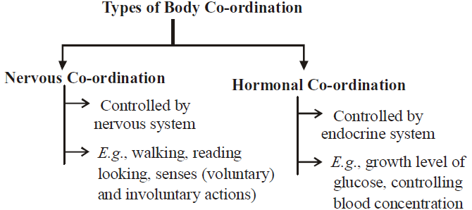
- In our body, the neural system and the endocrine system jointly coordinate and integrate all the activities of the organs so that they function in a synchronised fashion.
NEURAL SYSTEM
- A neural or nervous system is the system of neurons that form a network throughout the body for conducting information via electrical impulses so as to coordinate and control the activities of different parts as well as provide appropriate response to both internal and external stimuli.
- Stimulus is an agent, factor, chemical or change in external or internal environment which brings about a reaction in the organism.
- Response is the reaction of the organism to a stimulus.
- Receptors are cells, tissues and organs which are capable of receiving particular stimuli and initiate impulses to be picked up by sensory nerves.
- Impulse is self propagated electrical current that runs along the surface of the nerve fiber for passage of information.
- Effectors are muscles, glands, tissues or cells which act in response to a stimulus received from the nervous system.
- In Hydra and other cnidarians (coelenterates) like sea anemone and jellyfish, nervous system consists of several nerve cells linked with each other to form a sort of nerve nets in the body layer (epidermis and gastrodermis).
- The neural system is better organized in insects, where a brain is present along with a number of ganglia and neural tissues.
- In insects, nervous system consists of a bilobed nerve mass (the brain) present above the pharynx and the solid double nerve cord runs backward through the thorax and abdomen. It bears paired ganglia in the thorax as well as in abdomen.
- The vertebrates have a more developed neural system.
STRUCTURE OF NEURON OR NERVE CELL
- Neuron or nerve cell is a structural and functional unit of nervous system that is specialized to receive, conduct and transmit impulses. It is very long, sometimes reaching
90-100 cm. A neuron has three parts- cell body, dendrites and axon. The term neurites is used for both dendrites and axon.
CELL BODY OR CYTON
- It is broad, rounded, pyriform or stellate part of the neuron that contains a central nucleus, abundant cytoplasm and various cell organelles except centrioles. Because of the absence of centrioles, neurons cannot divide.
- Nucleus is large with a prominent nucleolus.
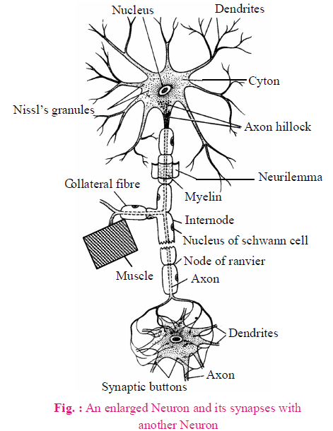
- Endoplasmic reticulum coils around the ribosome and form granule like structures called Nissl's granules or Tigroid body. It is the centre of protein synthesis. It is chemically ribonucleoprotein containing iron.
- Many small fibrils are found in the cytoplasm called neurofibrils, which help in internal conduction in the cyton.
DENDRON (DENDRITES)
- It is small cell process. It's fine branches called dendrites.
- Some receptors are found on the dendrites, so dendrons receive the stimuli and produce centripetal (towards the cell body) conduction.
- Dendrites contain Nissl’s granules and neurofibrils.
AXON
- It is the longest cell process of cyton, its diameter is uniform. It contains axoplasm.
- Nissl’s granules are absent in the axoplasm. It contains only neurofibrils and mitochondria.
- Axon is covered by axolemma. Part of cyton where axon arises is called axon hillock. The axon hillock is the neuron’s trigger zone, because it is the site where action potentials are triggered.
- The terminal end of axon is branched in button shape branches which are called as telodendria.
- More mitochondria are found in the telodendria which synthesize acetylcholine (Ach) with the help of choline acetyltransferase enzyme. Acetylcholine is stored in the synaptic vesicles.
- Axon is the functional part of nerve cell, therefore term nerve fibre usually refers to axon.
- Axon is covered by a layer of phospholipids (sphingomyelin) which is called as medulla or myelin sheath.
- Medulla is covered by thin cell membrane which is called as neurilemma or sheath of schwann cells.
- Schwann cells take part in the deposition of myelin sheath (myelinogenesis).
- Myelin sheath acts as an insulator and prevents leakage of ions and conserve axon’s energy.
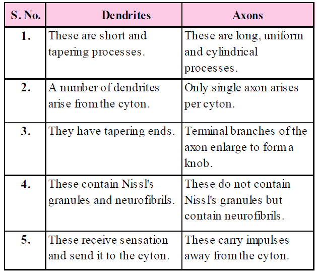
Table : Differences between Dendrites and Axons
TYPES OF NEURONS
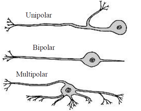
- Depending on the shape and the number and arrangement of the processes (axon and dendrites) arising from the cyton, three types of neurons are recognised:
- Unipolar neuron where the cyton is more or less spherical and has a single process that bifurcates. Such neurons are found usually in the embryonic stage.
- Bipolar neuron where the cyton is spindle shaped and has two processes, one at each end. Such neurons are found in the sense organs like ear and retina of the eye.
- Multipolar neuron where the cyton has several processes, one of which is long and forms the axon. Such neurons are found in the cerebral cortex.
- Apolar/Non-polar neuron : Cell processes are either absent or if present are not differentiated in axon and dendrons. Nerve impulse radiates in all directions, e.g., Hydra, cells of retina.
- Pseudo unipolar : In this type, cell has only axon but a small process develops from axon which act is as dendron, e.g., Dorsal root ganglia of spinal cord.
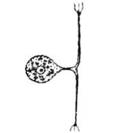
Fig. : Pseudo-Unipolar
- Depending on the function; three types of neurons are recognised :
- Sensory or Receptor neuron : These receive stimuli by their dendrites and transmit impulse towards central nervous system through their axon. These are found in sense organs.
- Motor or Effector neuron : The dendrites of these neurons synapse with axons of sensory neurons in the central nervous system. These send impulses from the central nervous system towards effectors, in response to stimuli.
- Relay or Connector neuron : These serve as links between sensory and motor neurons for distant relay of nerve impulses. These are found in central nervous system.
GENERATION AND CONDUCTION OF NERVE IMPULSE
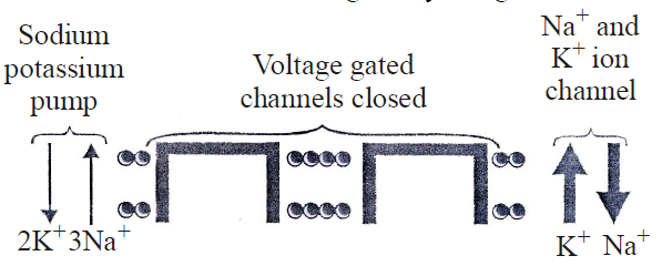
Neurons are excitable cells because their membranes are in a polarized state. Different types of ion channels are present on the neural membrane. These ion channels are selectively permeable to different ions.

RESTING MEMBRANE POTENTIAL IN RESTING PHASE
- The potential difference exists across the cell surface membrane of nerve cells. It is negative inside the cell with respect to the outside. This type of membrane is said to be polarised.
- The potential difference across the membrane at rest is called the resting membrane potential (RMP) and this is about – 70 mV (the negative sign indicates that inside the cell is negative with respect to the outside).
- The resting potential is maintained by active transport as well as passive diffusion of ions.
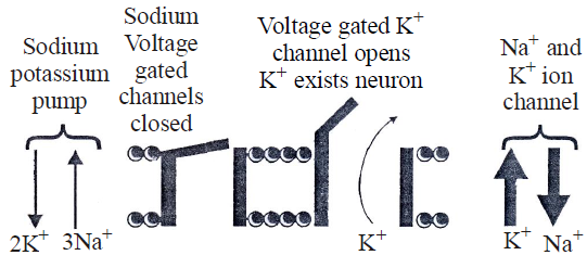
- Resting membrane potential is maintained by the active transport of ions against their electrochemical gradient by sodium potassium pump. These are carrier proteins located in the cell surface membrane. They are driven by the energy supplied by ATP and couple the removal of three sodium ions (3 Na+) from the axon with the uptake of two potassium ions (2K+).
- Because of this pump, the concentration of Na+ is more externally in the cytoplasm but K+ concentration becomes more in the axoplasm.
- The active movement of the ions is opposed by the passive diffusion of the ions. The rate of diffusion is determined by the permeability of the axon membrane to the ion. Potassium ions have a membrane permeability greater than that of sodium ions. Therefore, potassium ions loss from the axon is greater than sodium ion gain. This leads to a net loss of potassium ions from the axon, and the production of negative charge within the axon.
- Due to active transport (mainly) and diffusion process, positive charge is more outside and negative charge is more inside.
- Outer covering of axolemma is positively charged and inner membrane of axolemma is negatively charged.
ACTION POTENTIAL IN EXCITING STAGE
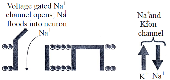
- Once the event of depolarization has occurred, a nerve impulse is initiated. Action potential is another name of nerve impulse. This is generated by a change in the sodium ion channels. These channels, and some of the potassium ion channels, are known as voltage gated channels, meaning these can be opened or closed with change in voltage. In resting state, these channels are closed due to binding of Ca++.
- An action potential is generated by a sudden opening of the sodium gates. Opening of gates increases the permeability of the axon membrane to sodium ions which enter by diffusion.
This increases the number of positive ions inside the axon. A change of –10mV in potential difference from RMP through influx is sufficiently significant to trigger a rapid influx of Na+ ions leading to generation of action potential. This change of –10 mV is called as threshold stimulus.
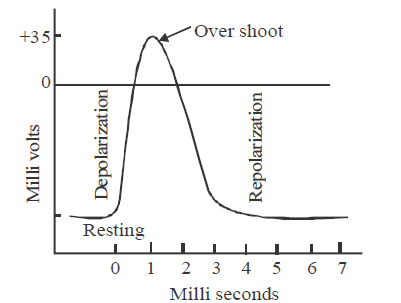
- At the point where membrane (axolemma) is completely depolarised due to rapid influx of Na+ ions, the negative potential is first cancelled out and becomes 0 (Depolarisation). This axolemma is called as excited membrane or depolarised membrane. Due to further entry of Na+, the membrane potential ‘‘over shoots’’ beyond zero and becomes positive upto +30 to +45mV. This ‘‘overshoot’’ peak corresponds to maximum concentration of sodium inside the axon. This potential is called the action potential. In this state, the inner surface of axolemma becomes positively charged and outer surface becomes negatively charged.
REPOLARISATION
- After a fraction of a second, the sodium gates close, depolarisation of the axon membrane causes potassium gates to open, potassium therefore diffuses out of the cell.
- Since potassium is positively charged, this makes the inside of cell less positive and activate Na+ – K+ pumps and the process of repolarization or return to the original resting potential begins.
- The repolarization period returns the cell to its resting potential (–70 mV). The neuron is now prepared to receive another stimulus and conduct it in the same manner.
- Sodium pump starts working to maintain the normal resting membrane potential by expelling Na+ and intaking of K+.
- The time taken for restoration of resting potential is called refractory period, because during this period the membrane is incapable of receiving another impulse.
- Nerve impulse travels as action potential which passes along the axon as a wave of depolarization.
- The whole process of depolarisation and repolarisation is very fast. It takes only about 1 to 5 milliseconds.
SALTATORY CONDUCTION OF NERVE IMPULSE
- This type of conduction occurs in myelinated fibre. Myelin is a fatty material with a high electrical resistance and act as an electrical insulator in the same way as the rubber and plastic covering of electrical wiring.
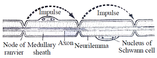
- The combined resistance of the axon membrane and myelin sheath is very high, but where breaks in the myelin sheath occur known as nodes of Ranvier, the resistance to current flow between the axoplasm and the fluid outside the cell is low. It is only at these nodes; local circuits are setup.
- This means, in effect that the action potential jump from node to node and passes along the myelinated axon faster as compared to the series of small local circuits in a non-myelinated axon. This type of conduction is called saltatory conduction. Leakage of ions takes place only in nodes of Ranvier and less energy is required for saltatory conduction.
SYNAPSE
- The term synapse was proposed by Charles Sherrington.
- It is the junctional region between two neurons where information is transferred from one neuron to another neuron but no protoplasmic connection.
Synapse = Pre synaptic knob + synaptic cleft + postsynaptic membrane
SYNAPTIC TRANSMISSION
- Neuron conducts the impulse in the form of electro-chemical wave.
- Conduction of nerve impulse is unidirectional.
- It follows "all or none" law. Magnitude of response will always be the same irrespective of strength of stimulus above threshold stimulus.
- Velocity of nerve impulse is directly proportional to the diameter of neuron.
- In mammals, the velocity of nerve impulse is 100 to 130 meter/sec.
- This velocity is affected by physical and chemical factors such as pressure, cold, heat, chloroform and ether etc.
- Telodendria of one neuron form synapse with dendron of next neuron.
- Telodendria membrane is called pre-synaptic membrane and membrane of dendron of other neuron is called as post-synaptic membrane. Space between pre and post-synaptic membranes is called synaptic cleft.
- Synapses are of two types - electrical synapse and chemical synapses.
- At electrical synapses, the membranes of pre- and post-synaptic neurons are very close by which electrical current easily flows from one neuron to the other. Impulse transmission across an electrical synapse is always faster than that across a chemical synapse. Electrical synapses are rare in our system.
- At chemical synapses, synaptic cleft is present. Chemicals called neurotransmitters are involved in the transmission of impulses at these synapses.
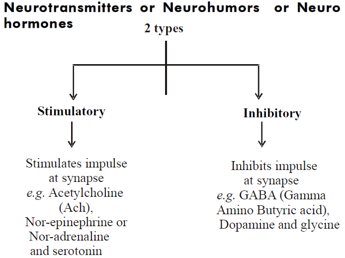
- When the action potential develops in pre synaptic membrane, it becomes permeable for Ca++. Ca++ enter in pre synaptic membrane & vesicles burst due to the stimulation of Ca++ and release of neurotransmitters (acetylcholine) in the synaptic cleft. Acetylcholine reaches the post-synaptic membrane via synaptic cleft and bind to receptors. It develops excitatory post-synaptic potential (EPSP). EPSP develop due to opening of Na+ gated channels.
- Cholinesterase enzyme is found in the synapse. This enzyme decomposes the acetylcholine into choline & acetate.
- Neuro-inhibitory transmitter Gamma Amino Butyric Acid (GABA) binds with post-synaptic membrane to open the
Cl– gated channels and hyperpolarization of neuron occurs. Now the potential is called inhibitory post-synaptic potential (IPSP) and further nerve conduction is blocked.
Table : Differences between Electrical and Chemical Synapses
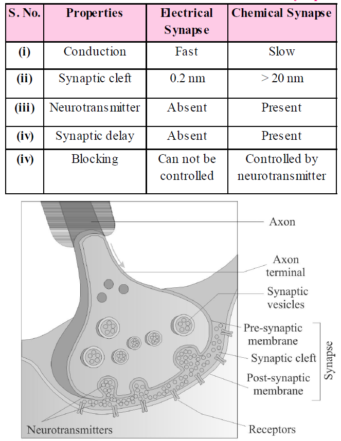
Fig. : Diagram showing axon terminal and synapse
Neuroglia/Glial cells : These are supporting cells which form a packing substance around the neurons.
NERVOUS SYSTEM
- It is the system which regulates the various activities of the body through nerve-impulses by which the stimulus are transmitted at a faster rate.
- The nervous system controls and coordinates the various activities of the organs of the animals.
- Whole nervous system of human being is derived from embryonic ectoderm.
CENTRAL NERVOUS SYSTEM
- The brain is the central information processing organ of our body, and acts as the ‘command and control system’. It controls the voluntary movements, balance of the body, functioning of vital involuntary organs (e.g., lungs, heart, kidneys, etc.), thermoregulation, hunger and thirst, circadian (24-hour) rhythms of our body, activities of several endocrine glands and human behaviour. It is also the site for processing of vision, hearing, speech, memory, intelligence, emotions and thoughts.
- It includes the brain and the spinal cord. These are formed from the neural tube which develops from the ectoderm after the gastrula stage of the embryo.
- Development of CNS : It develops from neural tube. Anterior part of neural tube develops into brain while caudal part of neural tube develops into spinal cord. Approximately 70-80% part of brain develops in 2 years of age & complete development is achieved in 6 years of age & spinal cord develops completely in 4 to 5 years of age.
BRAIN
It is situated in cranial box of skull which is made up of 8 bones i.e., 1 frontal bone, 2 parietal bones, 2 temporal bones, 1 occipital bone, 1 ethmoid bone, 1 sphenoid bone. The weight of the brain of an adult man is 1400 gm and of female is 1250 gm.
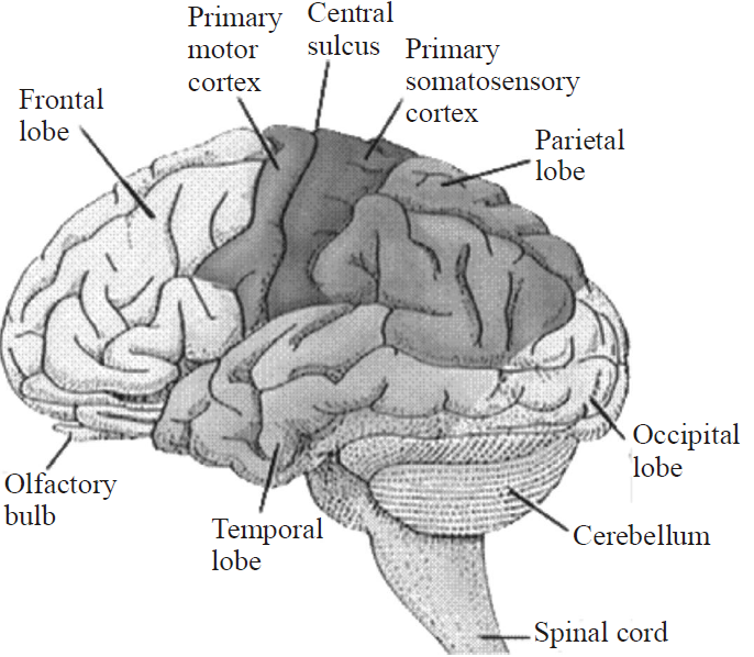
Fig. : Diagram showing section of the human brain
BRAIN MENINGES
Brain is covered by three membranes of connective tissue termed as meninges or menix.
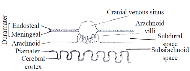
Fig. : Meningel layer
(1) Dura mater
- This is the first and the outermost membrane which is thick, very strong and non-elastic. It is made up of collagen fibres. This membrane is attached with the innermost surface of the cranium.
- It is double layer-outer endosteal layer which is closely attached with inner most surface of cranium & no space is found between skull and duramater (no epidural space). Inner meningeal layer which is related with other meninges of brain, both are vascular. Generally both layers are fused with each other, but at some places these are separated from one another & form a sinus called cranial venous sinus. These sinuses are filled with venous blood.
(2) Arachnoid
It is middle, thin and delicate membrane. It is found only in mammals. It is non vascular layer. In front of cranial venous sinus, it becomes folded, these folds are called arachnoid villi. These villi reabsorb the cerebrospinal fluid (CSF) from sub arachnoid space & pour it into cranial venous sinuses.
(3) Pia mater
- It is innermost, thin and transparent membrane, made up of connective tissue. Dense network of blood capillaries are found in it, so it is highly vascular.
- It is firmly adhered to the brain. Pia mater & arachnoid layer at some places fuse together to form leptomeninges. Pia mater merges into sulci of brain & densely adhere to it. At some places, it directly merges in the brain and called telachoroidea
- Telachoroidea form the choroid plexus in the ventricles of brain.
SUB DURAL SPACE
Space between the dura mater & arachnoid. It is filled with serous fluid.
SUB ARACHNOID SPACE
Space between arachnoid & pia mater is filled with cerebro-spinal fluid. S.F. cranial nerves also pass through this space.
MENINGITIS
Any inflammation of meninx is called meningitis. It may be caused by viruses, bacteria or protozoa.
CEREBROSPINAL-FLUID (CSF)
- This fluid is clear and alkaline in nature just like lymph. It has protein (albumin, globulin), glucose, cholesterol, urea, bicarbonates, sulphates and chlorides of Na, K. Protein & cholesterol concentration is lesser than plasma & Cl– conc. is higher than plasma.
- In a healthy man, in 24 hrs, 500 ml of cerebrospinal fluid is formed & absorbed by arachnoid villi. At a time, total volume of cerebrospinal fluid is 150 ml.
- Cerebrospinal fluid is present in ventricle of brain, sub-arachnoid space of brain & spinal cord.
Functions of CSF
- It acts as a shock absorbing medium and work as a cushion for protection of brain.
- It provides buoyancy to the brain, so net weight of the brain is reduced from about 1.4 kg to about 0.18 kg.
- Excretion of waste products.
- Endocrine medium for the brain to transport hormones to different areas of the brain.
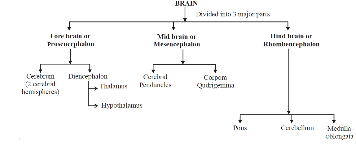
(1) FOREBRAIN
The forebrain consists of cerebrum, thalamus and hypothalamus.
(a) Cerebrum
- It is the first and most developed part of the brain. It makes 2/3 part of total brain.
- Cerebrum consists of two cerebral hemispheres on the dorsal surface. Many ridges and grooves are found on the dorsal surface of cerebral hemisphere. Ridges are known as gyri while grooves are called sulci. These cover the 2/3rd part of cerebrum. Gyri and sulci are more developed in human beings so human beings are the most intelligent living beings. A longitudinal groove is present between two cerebral hemispheres called as median fissure. Both the cerebral hemispheres are partially connected with each other by curved thick nerve fibres is called the corpus callosum.
- The layers of cells which covers the cerebral hemisphere is called the cerebral cortex and is thrown into prominent folds. It is referred to as the grey matter due to its grayish appearance. Inner to it is cerebral medulla of white matter. Grey matter is made of cell bodies while white matter is formed of myelinated nerve fibers.
- The cerebral cortex contains motor areas, sensory areas and large regions that are neither clearly sensory nor motor function. These region called as the association areas which are responsible for complex functions like intersensory associations, memory and communication.
- Each cerebral hemisphere is divided into five parts— frontal, parietal, temporal, occipital and insula .
- Insula Lobes : These are hidden due to being present in the deep sylvian fissure.
- Frontal Lobes : These occur in front or anterior region. Frontal lobes are centres of intelligence. These control various types of movements (both voluntary and involuntary) including facial muscles, chewing, swallowing, movement of tongue, movement of lips, etc.
- Parietal Lobes : These are situated in the mid upper area. Parietal lobes have centres for taste, senses (sensations of pain, touch, pressure, and temperature) and some components of speech.
- Temporal Lobes : Temporal lobes control hearing, smell, recall of audio-visual events and some components of speech.
- Occipital Lobes : Occipital lobes have centres of perception of sight.
(b) Diencephalon
- It is small and posterior part of fore forebrain. It is covered by cerebrum. Its dorsal side is called epithalamus in which pineal body is situated, that controls the sexual maturity of animal.
- It consists of the thalamus and hypothalamus.
- Thalamus
- It forms upper lateral walls of diencephalon. It forms 80% part of the diencephalon.
- It acts as a relay centre. It receives all sensory impulses from all parts of body & these impulses are sent to the cerebral cortex.
- Hypothalamus
- It forms lower lateral wall of the diencephalon.
- A cross like structure is found on the anterior surface of the hypothalamus called the optic chiasma. It carries optic impulses received from eyes to the cerebral hemispheres. Animal becomes blind if this part is destroyed by chance.
- Pituitary body is attached to the middle part of hypothalamus by the infundibulum.
- Corpus mamillare or Corpus albicans is found on the posterior part of the hypothalamus. It is a character of mammalian brain.
- Hypothalamus has control centers for hunger, thirst, fatigue, sleep, sweating, body temperature and emotions. It also secretes a number of hormones.
- The inner parts of cerebral hemispheres and a group of associated deep structures like the amygdala, hippocampus, etc., form a complex structure called the limbic lobe or limbic system. Along with the hypothalamus, it is involved in the regulation of sexual behaviour, expression of emotional reactions (e.g., excitement, pleasure, rage and fear), and motivation.
(2) MIDBRAIN
- It is located between the thalamus/hypothalamus of the forebrain and pons of the hindbrain.
- Anterior part of midbrain contains two longitudinal myelinated nerve fibres peduncles called cerebral peduncles or crura cerebri.
- Crura cerebri controls the muscles of limbs.
- On the posterior part of midbrain are found four spherical projections called colliculus or optic lobes. Four colliculus are collectively called as corpora quadrigemina (2 upper and 2 lower).
(3) HINDBRAIN
It comprises pons, cerebellum and medulla oblongata.
(a) Pons or Pons varolii
- It is a small spherical projection, which is situated below the midbrain and upper side of the medulla oblongata.
- Pons function as relay centre among different parts of the brain. It also possesses pneumotaxic area of respiratory centre.
(b) Cerebellum
- It is made up of 3 lobes [2 lateral lobes and 1 vermis (divide in 9 segments)].
- It is the second largest part of the brain, constituting about 12.5% of the total volume of brain. It lies behind the cerebrum and above the medulla oblongata.
- It coordinates muscular activity of the body and also maintains equilibrium or posture of the body as during walking, jumping, lifting, catching, bending, etc.
(c) Medulla Oblongata
- It is the hindermost part of the brain which lies below cerebellum. It continues behind into spinal cord. Medulla oblongata has fluid filled cavity called fourth ventricle. Its roof bears posterior choroid plexus (for filtering cerebrospinal fluid from blood) and three pores for connecting external cerebrospinal fluid with internal cerebrospinal fluid. Medulla oblongata contains –
- Respiratory centre for regulating the rate of breathing.
- Cardiac centre for regulating the rate of heart beat.
- Regulation of blood pressure.
- Reflex centre for swallowing, vomiting, coughing, sneezing, salivation etc.
- Pons, medulla oblongata and midbrain are collectively called brain stem.
INTERNAL STRUCTURE OF BRAIN
- One pair of small, spherical and solid olfactory lobes are present in the human brain. No ventricle is found in it. Both olfactory lobe are separate from each other & are embedded into ventral surface of both frontal lobe of cerebral hemisphere.
- Olfactory lobe is supposed to be the centre of smelling power. Its size is small in mammals comparatively because most of its parts become a part of cerebrum. Some animals like sharks and dogs have well developed olfactory lobes.
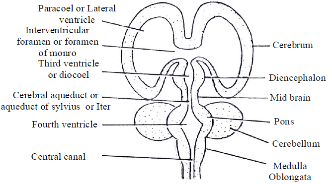
Fig. : L.S. of Brain
- Except midbrain, cerebellum, pons & olfactory lobe, complete brain is internally hollow. Its cavity is lined by ependymal epithelium (ciliated columnar epithelium).
- Cavities of brain are known as ventricles, filled with cerebrospinal fluid (CSF).
- Function : Formation of CSF by secretion of plasma.
Basal Ganglia
- These consist of two main structures, the corpus striatum (the biggest nucleus of basal ganglia) and red nucleus.
- Corpus striatum is further differentiated into Lenticular nucleus and Caudate nucleus.
- The Lenticular nucleus contains 2-parts, the pallidum and putaman.
- Functions
- It maintains muscle tone.
- It regulates automatic associated movements like swinging of arms during walking.
- In lower animals, when cerebral cortex is not developed basal nuclei acts as motor centre.
- Parkinson’s disease (shaking palsy) develops due to the deficiency of a neurotransmitter (Dopamine) in a part of the basal ganglia.
Limbic System
- It is visible like a wishbone, tuning fork or lip like neural link between cerebrum and brainstem.
- It includes limbic lobes (area of temporal lobe), hippocampus, hypothalamus including septum, part of thalamus and amygdaloid complex.
- Functions of Limbic System
- It converts behaviour, emotion, rage and anger (hypothalamus, amygdaloid body).
- It converts recent memory and short term memory into long term memory. (Hippocampal lobe).
FUNCTIONS OF BRAIN
- Sensory Information : Brain receives information from all the sensory receptors and sense organs of the body.
- Processing : It processes the information obtained from various sources and chooses the most appropriate response.
- Response : Brain sends instructions to effector organs all over the body to provide the appropriate response to received stimuli.
- Control : It has controls for regulating the functioning of various body organs.
- Coordination : Working of the different organs of a system is coordinated by the brain.
- Reflexes : It has centres for reflexes related to sound, sight and involuntary functioning of many body parts.
SPINAL CORD
- The spinal cord is a long, thin tubular bundle of nervous tissue and support cells that extends from the brain (the medulla oblongata specifically).
- The spinal cord begins at the occipital bone and extends down to the space between the first and second lumbar vertebrae; it does not extend the entire length of the vertebral column.
- Its upper part is wide while lowermost part is narrow known as conus-medullaris.
- Conus medullaris is present upto L1 vertebra. Terminal part of conus medullaris extend in the form of thread like structure made up of fibrous connective tissue called filum terminale.
- The spinal cord is protected by 3 layers of tissue, called spinal meninges, that surround the canal.
- The dura mater is the outermost layer, and it forms a tough protective coating. Between the dura mater and the surrounding bone of the vertebrae is a space called the epidural space.
- The arachnoid mater is the middle protective layer.
- The pia mater is the innermost protective layer. It is very delicate and it is tightly associated with the surface of the spinal cord.
- The outer-part of spinal cord is of white matter while inner-part contains grey matter.
- On the dorso-lateral & ventro-lateral surface of spinal cord, the gray matter projects outside & forms the one pair dorsal & ventral horn.
- Due to formation of dorsal & ventral horn, white matter is divided into 4 segments and segment is known as funiculus or white column.
- The spinal cord has 3 major functions : as a conduit for motor information which travels down the spinal cord, as a conduit for sensory information in the reverse direction and finally as a center for coordinating certain reflex.
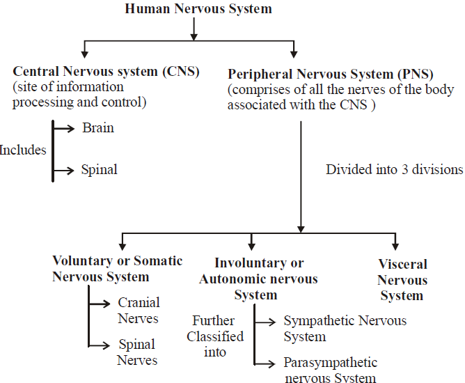
PERIPHERAL NERVOUS SYSTEM
- All the nerves arising from the brain and spinal cord are included in peripheral nervous system.
- Nerves arising from the brain are called cranial nerves, and nerves coming out of the spinal cord are called spinal nerves.
- The nerve fibres of the PNS are of two types :
- Afferent fibres : These transmit impulses from tissues/organs to the central nervous system (CNS).
- Efferent fibres : These transmit regulatory impulses from the CNS to the concerned peripheral tissues/organs.
- The peripheral nervous system (PNS) is divided into 3 divisions –
- Somatic nervous system (SoNS) or Voluntary nervous system
- Autonomic nervous system (ANS)
- Visceral nervous system (VNS)
SOMATIC NERVOUS SYSTEM
It is associated with the voluntary control of body movements via skeletal muscles. The SoNS consists of efferent nerves responsible for stimulating muscle contraction, including all the non-sensory neurons connected with skeletal muscles and skin.
It consists of three parts :
- Cranial nerves
- Spinal nerves
- Association Nerves
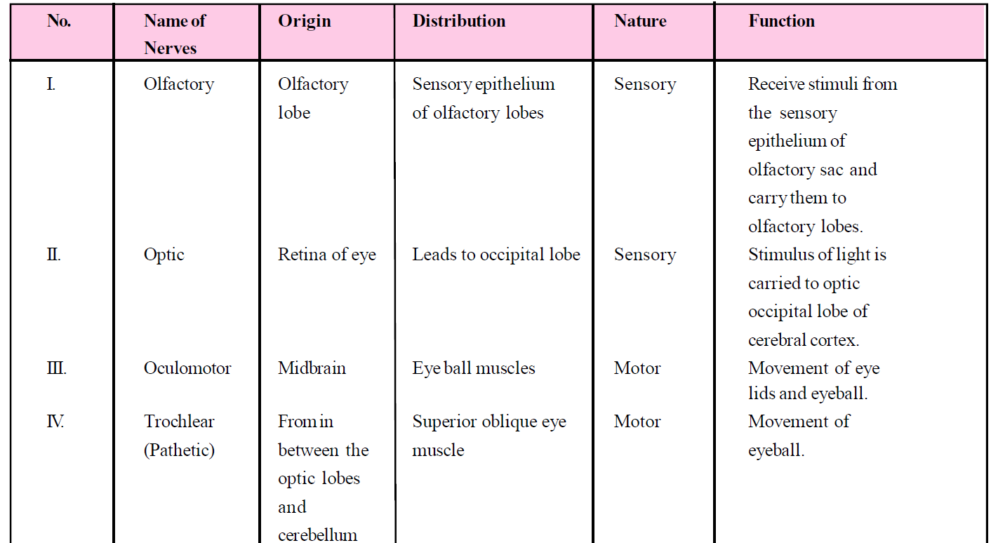
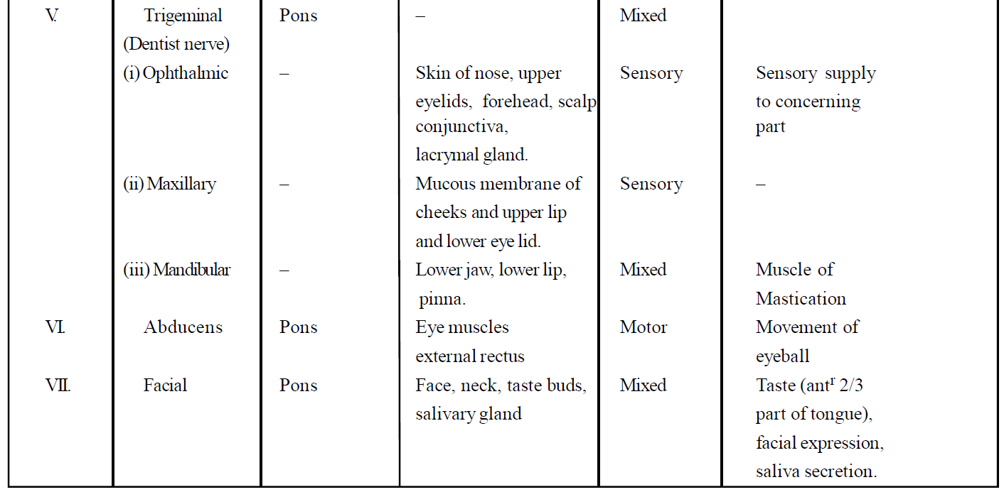
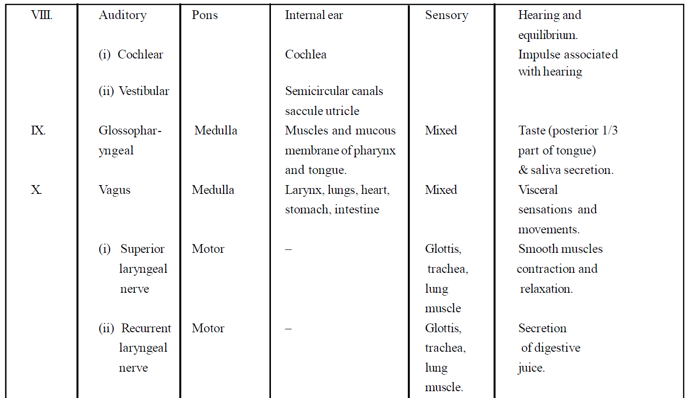
(1) CRANIAL NERVES
- 12-pairs of cranial nerves are found in reptiles, birds and mammals but amphibians and fishes have only 10 pairs of cranial nerves.
- In human, I, II and VIII are cranial nerves out of 12 pairs of total, cranial nerves are pure sensory in nature.
- III, IV, VI, XI and XII cranial nerves are motor nerves and V, VII, IX & X cranial nerves are mixed type of nerve.
- Fibres of autonomous nervous system are found in III, VII, IX & X cranial nerves.
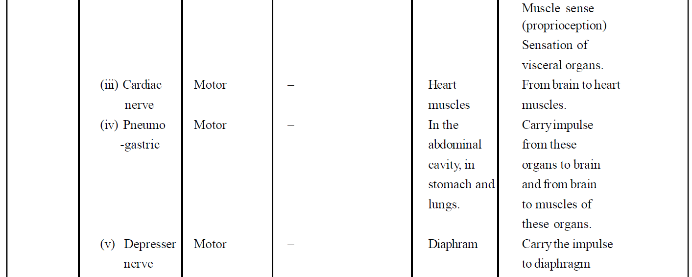
- Longest cranial nerve is Vagus nerve.
- Largest cranial nerve is Trigeminal nerve.
- Smallest cranial nerve is Abducens nerve.
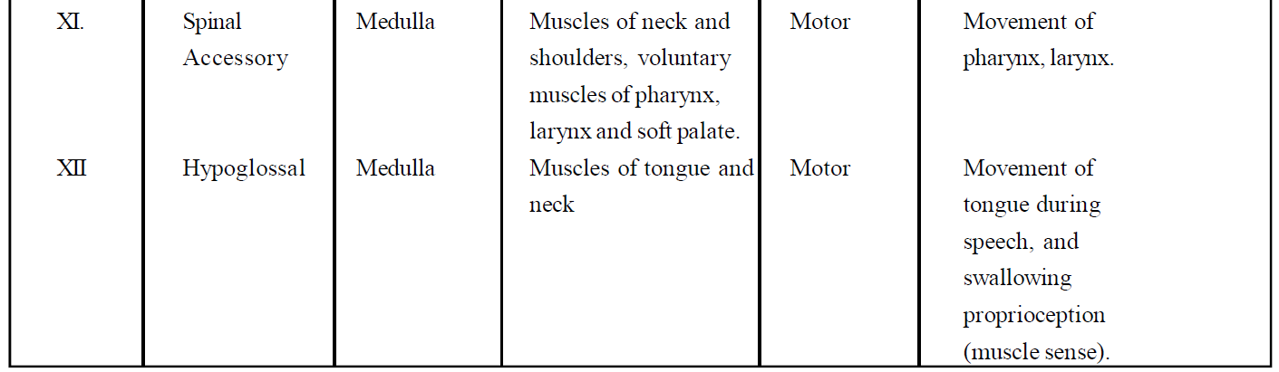
Table : Summary of Human Cranial Nerves
(2) SPINAL NERVES
- In rabbit, there are 37 pairs of spinal nerves, while in frog there are 9 or 10 pairs of spinal nerves.
- In human, only 31 pairs of spinal nerves are found.
- The spinal nerves in man are divided into 5 groups.
(1) Cervical (C) → 8 pairs — in neck region
(2) Thoracic (T) → 12 pairs — in thoracic region
(3) Lumbar (L) → 05 pairs — upper part of abdomen
(4) Sacral (S) → 05 pairs — lower part of abdomen
(5) Coccygeal (CO) → 01 pair — represent the tail nerves.
Total = 31 pairs
- Each spinal nerve is mixed type and arises from the roots of the horns of grey matter of the spinal cord. In dorsal root, only afferent or sensory fibres and in ventral root efferent or motor fibres are found.
- Both the roots after moving for distance in the spinal cord of vertebrates combine with each other and come out from the intervertebral foramen in the form of spinal nerves.
(3) ASSOCIATION NERVES
These nerves integrate sensory input and motor output.
AUTONOMIC NERVOUS SYSTEM
- The autonomic nervous system is that part of the peripheral nervous system which controls activities inside the body that are normally involuntary, such as heartbeat, gut peristalsis, sweating etc.
- It consists of motor neurons passing to the smooth muscles of internal organs. Smooth muscles are involuntary muscles. Most of the activities of the autonomic nervous system is controlled within the spinal cord or brain by reflexes known as visceral reflexes and does not involve the conscious control of higher centres of the brain.
- Overall control of the autonomic nervous system is maintained, however, by centres in the medulla (a part of the hindbrain) and hypothalamus.
- These receive and integrate sensory information and coordinate this with information from other parts of the nervous system to produce the appropriate response.
- ANS plays an important role in maintaining the constant internal environment (homeostasis).
- There are two divisions of the autonomic nervous system the sympathetic (SNS) nervous system and parasympathetic (PNS) nervous system.
- Sympathetic and parasympathetic divisions typically function in opposition to each other. But this opposition is better termed complementary in nature rather than antagonistic. The sympathetic division typically functions in actions requiring quick responses. The parasympathetic division functions with actions that do not require immediate reaction. Consider sympathetic as "fight or flight" and parasympathetic as "rest and digest" or "feed and breed".
Nervous Control of Visceral Organs
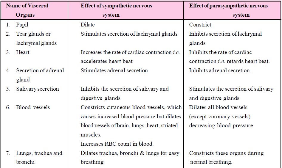
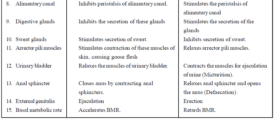
VISCERAL NERVOUS SYSTEM
Visceral nervous system is the part of the peripheral nervous system that comprises the whole complex of nerves, fibers, ganglia and plexuses by which impulses travel from the central nervous system to the viscera and from the viscera to the central nervous system.
REFLEX ACTION
- Marshal Hall first observed the reflex actions.
- Reflex actions are spontaneous, automatic, involuntary, mechanical responses produced by specific stimulating receptors.
- It is a form of animal behaviour in which the stimulation of a sensory organ (receptor) results in the activity of some organs without the intervention of will.
- Reflex actions are of 2 types:
- Cranial reflex : These actions are completed by brain. No urgency is required for these actions. These are slow actions, e.g., watering of mouth to see good food.
- Spinal reflex : These actions are completed by spinal cord. Urgency is required for these actions. These are very fast actions, e.g., displacement of the leg at the time of pinching by any needle.
- Classification of reflex actions on the basis of previous experiences:
- Conditioned reflex : Previous experience is required to complete these actions e.g., swimming, cycling, dancing, singing etc. These actions were studied first by Evan Pavlov on dog. Initially these actions are voluntary at the time of learning and after perfection these become involuntary.
- Unconditioned reflex : These actions do not require previous experience, e.g., sneezing, coughing, yawning, sexual behaviour for opposite sex partner, migration in birds etc.
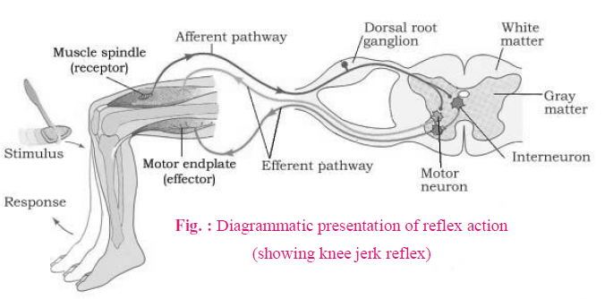
REFLEX ARC
The path of completion of reflex action is called reflex arc.
Sensory fibres carry sensory impulses in the grey matter. These sensory impulses are converted now into motor impulses and reach up to muscles. These muscles show reflex actions for motor impulses obtained from motor neurons. Reflex arc is of two types :
- Monosynaptic : In this type of reflex arc, there occurs direct synapse (relation) between sensory and motor neurons. Thus, nerve impulse travels through only one synapse, e.g., Stretch reflex
- Polysynaptic : In this type of reflex arc, there are one or more small neurons in between the sensory and motor neurons. These small neurons are called connector or interneurons, e.g., withdrawal reflex. Nerve impulse will have to travel through more than one synapses in this reflex arc.

SENSORY RECEPTION AND PROCESSING
- A sensory system is a part of the nervous system responsible for processing sensory information. A sensory system consists of sensory receptors, neural pathways, and parts of the brain involved in sensory perception.
- We smell things by our nose, taste by tongue, hear by ear and see objects by eyes.
- The sense receptors on the tongue and within the nasal cavity work very closely together to give us our sense of taste. These 5 kinds of receptors-the olfactory cells in the nose and the four special cells or taste buds (gustatory receptors) on the tongue for discriminating salty, sweet, sour, and bitter tastes also have a functional similarity. The neurons of the olfactory epithelium extend from the outside environment directly into a pair of broad bean-sized organs, called the olfactory bulb, which are extensions of the brain's limbic system.
HUMAN EYE
- It is a photoreceptor organ (photoreceptor part-retina)
- It is ecto - mesodermal in origin.
- The wall of eyeball is composed of three layers; i.e., scleroid, choroid and retina.
SCLEROID
It consists of white fibrous connective tissue, and therefore, looks white. The anterior one sixth part of the eyeball, visible externally, is transparent and is called the cornea. The major part (5/6) of the eyeball is white.
CHOROID (UVEA)
- It is pigmented and highly vascularised coating.
- Unlike scleroid, it is incomplete in the anterior region, and forms ciliary bodies and iris.
- The pupil (an opening for light entry) is present in the centre of the iris. The eye colour is the colour of the iris.
- Ciliary body secretes aqueous humour which provides nourishment to the lens and cornea because these do not have the blood supply of their own. The 2-chambers which contain aqueous humour are anterior (between the cornea and iris) and posterior (between iris and lens) chambers.
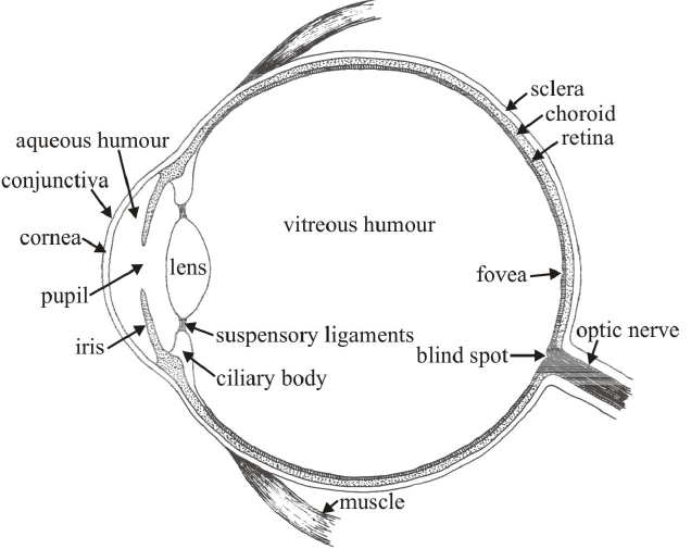
Fig. : V. S. of mammalian eye
LENS
- The lens in human eye is biconvex, circular, living, multi-cellular and non-vascularised (no blood supply).
- It is purely ectodermal in origin.
- Position of the lens in human is fixed but the focal length is adjustable. This adjustment of focal length by thinning or thickening of the lens is called accommodation power.
- The focal length (f) of human eye lens is 1.5 cm, with refractive power of 66.7 Diopter at rest.
RETINA
- It is sensory coating of the eye.
- In human eye, the retina is inverted (sensory cells lie opposite to the entry of light).
- From outer to inner, the retina has 4 prominent layers –
(i) Pigmented epithelium (ii) Sensory layer (iii) Bipolar neurons-layer (iv) Ganglionic layer with optic nerve fibres.
- The pigmented epithelium absorbs oblique (scattered) light rays to prevent internal reflection (on the retina).
- The sensory layer has two types of sensory cells –
(i) Rods (ii) Cones
- In each human eye, the number of rods (~120 million) is 20 times of that of cones.
- The yellow spot at the visual axis of retina is called the macula lutea. This contains only cones (rods absent). The sharpest vision occurs at the central concave point of the yellow spot called fovea centralis.
- The point from where optic nerve arises is called blind spot. Both rods and cones are absent at this point and therefore, there is no vision.
- The rods are sensitive for vision in dim light (for black & white vision). The night vision (i.e., vision in dim light) is also called ‘scotopic vision’.
- The pigment present in rods is rhodopsin (visual purple).
- The cones are sensitive to bright light (for colour vision). The colour vision is called ‘photopic vision’.
- The pigment present in cones is called photopsin (visual violet).
- Between retina and lens there is a jelly-like mucous connective tissue, called vitreous humour. It is permanent refractive medium and maintains the shape of the eyeball.
- The image formed on the retina of human eye is laterally inverted and reversed (upside down).
MECHANISM OF VISION
- The light rays from the object pass through the conjunctiva, cornea, aqueous humour, lens and vitreous humour in that order. All these structures refract the light such that it falls on the retina. This is called focussing. Maximum focussing is done by the cornea and the lens. The light then falls on the retina.
- This light is received by the photoreceptors-rods and cones on the retina. The absorbed light activates the pigments present in the rods and cones. The pigments are present on the membranes of the vesicles. Thus, the light is then converted into action potentials in the membranes of the vesicles. These travel as nervous impulses through the rod or cone cell and reach the synaptic knobs. From here the impulses are transmitted to the bipolar nerve cells, then to the ganglions and then to the optic nerves. Thus, the nervous impulses generated in the retina are carried to the brain by about a million neurons of the optic nerve. The vision is controlled by the occipital lobe at the back of the brain. The information received is processed and we are able to see the image. The image formed on the retina is inverted. However, the brain makes us see the image erect. So, through the eyes are essential for vision, any damage to the optic nerves also results in impairment of vision.
REFRACTIVE ERROR OF EYE
- Myopia : Image form in front of retina and far objects are not clearly seen.
Reason - The increased curvature of the cornea or increase convexity of lens.
Prevention - By biconcave (diverging) lens myopia can be corrected.
- Hypermetropia : Image is formed behind the retina. So near objects are not seen clearly.
Reason - In this, lens become flat or decreased curvature of cornea decreases convexity of lens. Eye of new born baby is "hypermetropic".
Prevention - It is corrected by biconvex (converging) lens.
- Presbiopia : After 35 to 40 years, elasticity of lens decrease, so hypermetropia occur. Hypermetropia due to ageing is called presbyopia. Corrected by biconvex lens.
- Astigmatism : Due to different curvature of lens at the different place overall image is not form at the yellow spot. Cylindrical lens are used for the correction of this disorder.
OTHER DISEASES
- Cataract : After 60 years, lens become opaque due to destruction of cysteine and glutathione amino acids. In India, it is the most common cause of blindness (80%).
Treatment : Removal of cataractal lens and replace by intraocular lens.
- Glaucoma : At the junction of cornea and sclera, there is a canal called schlemm's canal. This canal drains out aqueous humour into the veins. Aqueous humour is formed by the blood vessels of ciliary process. Sometimes Schlemm canal gets blocked, so drainage of aqueous humour does not occur. Hence, aqueous humour gets collected in the anterior and posterior chamber of eye. So IOP (Intraocular pressure) increases (normally 16-23 mm of Hg) and condition is called Glaucoma.
- Trachoma : It is an infectious disease caused by Chlamy-dia trachomatis bacterium which produced a characteristic roughening of the inner surface of the eyelids. Occurring of inflammation of conjunctiva called conjunctivitis. Eyes become red.
HUMAN EAR
It is a statoacoustic organ, i.e., for balancing as well as hearing.
Human ear has 3 parts –
- External ear – Pinna + Auditory meatus
- Middle ear – Tympanic cavity + ear ossicles
- Internal ear – Vestibular apparatus + cochlea.
Between external and middle ear, an ear drum (Tympanum) is present. Similarly, between middle ear and internal ear, there are two membrane-bound windows – Oval window (Fenestra ovalis) and Round window (Fenestra rotundus) are present.
(1) EXTERNAL EAR
- Pinna (Auricula), a characteristic of mammals, directs sound vibrations towards auditory meatus. It is made up of elastic cartilage.
- Ceruminous glands are present in the lining of auditory meatus and secrete ear wax (Cerumen). The ear wax is sticky and prevents the entry of dust particles to the tympanum. It also prevents fungal and bacterial infections.
(2) MIDDLE EAR
- The cavity of the middle ear is called tympanic cavity. It develops from pharyngeal out-growth and is, therefore, endodermal in origin.
- Eustachian tubes connect middle ear to pharynx and prevent the rupturing of tympanum during louder sound by equalizing the pressure on the back of the membrane. The Eustachian tubes open during yawning, swallowing and chewing.
- The tympanic cavity contains 3-ear ossicles (smaller bones). From the tympanum to the oval window, the sequence of these bones is –
- Malleus – Hammer shaped
- Incus – Anvil shaped
- Stapes – Stirrup shaped
- The ear ossicles not only conduct sound vibrations but also amplify them.
Stapes, the smallest bone of mammalian body, fits on to the oval window.
(3) INTERNAL EAR
- The oval window is the inlet for the sound vibrations. Because of the smaller size of oval window the pressure of sound is also amplified on this membrane by about 20 times of the pressure on tympanum.
- The sensory part of internal ear is called membranous labyrinth. It is filled with
Endolymph. - The membranous labyrinth is surrounded by bony labyrinth, formed by ‘Temporal bone’. Between membranous labyrinth and bony labyrinth, the fluid is Perilymph.
- The internal ear has two prominent parts –
(a) Vestibular Apparatus
- It is sensory part for balancing the body. It is also well developed in fishes.
- It consists of 3-semicircular canals, one utriculus and one sacculus.
- Out of 3-semicircular canals (SCC), two are vertical and one horizontal.
- Each SCC is surrounded by bony canal. The fluid in between bony and membranous canal, as mentioned earlier, is perilymph.
- At one end of each canal, there is an ampulla.The ampulla has sensory hair cells which are bathed in endolymph.
- Utriculus also has a group of ‘hair cells’ at its floor, called Macula Utriculus (MU) which is bathed in endolymph.
- The hair cells of MU are surrounded by an otolithic membrane in which calcium carbonate crystals (otoconia/otolith) are embedded.
- Sacculus too contains group of hair cells lying in the walls in semi-vertical position. This group of hair cells is known as Macula sacculus (MS).
(b) Cochlea
- It is sensory structure for hearing.
- It is a coiled structure, having 2 ¾ coils in human.
- The cochlea, in cross section, shows 3-canals.
- Vestibular canal (Scala Vestibuli)
- Cochlear (median) canal (Scala Media)
- Tympanic canal (Scala Tympani)
- Vestibular canal and tympanic canal are filled with perilymph while cochlear canal is filled with endolymph. The cochlea, therefore contains both perilymph and endolymph fluids. The cochlear or median canal, filled with endolymph, is the actual part of the membranous labyrinth.
- The group of sensory ‘hair cells’ for hearing are present in cochlear canal or scala media.
- The membrane at the roof of scala media which separates scala vestibuli from scala media is called Reissner membrane. The membrane at the floor of scala media, which separates scala media from scala tympani is called Basilar membrane.
- The ‘hair cells’ are attached to the basilar membrane and form ‘Organ of Corti’, the ultimate sensory part for hearing. These hair cells are supported by Dieter cells. The group of hair cells is covered by a secretory (non-cellular) membrane called tectorial membrane, into which the processes of hair cells are embedded.
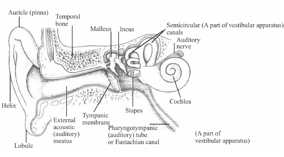
Fig. : Structure of Ear
MECHANISM OF HEARING
- Pinna collects and amplifies sound waves which then pass along the auditory canal to the eardrum.
- Sound waves strike the eardrum and cause vibrations in the thin stretched membrane. The eustachian tube equalizes air pressure on either side of the eardrum which allows a free vibration.
- The vibration reaches ear ossicles in the middle ear. Ear ossicles transmit vibrations from the air to the denser fluid in the ear.
- The lever-like action of malleus and incus magnifies the vibration of the stapes.
- The vibrating stapes transmits vibrations to the membrane of the oval window. Round window reduces the movement of fluid in the cochlea.
- Vibrations from oval window transmit to cochlea. This leads to vibration of the fluid in the cochlear canals.
- Vibrations of fluid in cochlear canals triggers movement of hair cells of the organ of corti in cochlea.
- Movement of hair cells is converted to a nerve signal.
- Nerve signal is transmitted to the brain via the auditory nerve and this results in hearing.
Study Notes for NEET/AIIMS/JIPMER


