


LOCOMOTION AND MOVEMENT
INTRODUCTION
Locomotion is the ability to move in a particular direction in its environment, which requires a propulsive force acting against a supporting structure.
TYPES OF MOVEMENTS
- Movement of body parts is performed by all living organisms including plants.
- Animals are able to move their body parts by movement of external body parts or internal organs.
- Purpose of external body part movements are
- To collect information.
- Maintenance of equilibrium.
- Locomotion.
- Capturing and ingestion of food.
- Breathing.
- Purpose of internal body parts movements are
- Circulation of blood.
- Gaseous exchange.
- Peristalsis movement for passage of food and urine.
- Movements of gametes and foetus in genital tract and uterus during fertilization and child birth respectively.
- Human body exhibit three main types of movements: amoeboid, ciliary and muscular.
- Amoeboid movement occurs by means of temporary protoplasmic outgrowth (called pseudopodia) which are formed due to flow of protoplasm in any direction. E.g., Amoeba, macrophages and leukocytes. Cytoskeletal elements like microfilaments are also involved in amoeboid movement.
- Ciliary movement occurs in most of our internal tubular organs which are lined by ciliated epithelium. Passage of ova through the female reproductive tract is also facilitated by the ciliary movement.
- Muscular movements is caused by the property of contractility of muscles, which is used effectively for locomotion. Movement of limbs, jaws, tongue etc. require muscular movement.
TYPES OF LOCOMOTION
- Locomotion requires a perfect coordinated activity of muscular, skeletal and neural systems.
- Locomotion takes several forms such as walking (man), creeping (earthworm, lizard), cursorial (horse, flightless birds), hopping (frog, rabbit), running (dog, horse), flying (insects, birds) and swimming (fish, whale).
LOCOMOTION IN DIFFERENT ANIMALS
- Protozoa : Locomotion in protozoans occurs by the help of cilia, flagella and pseudopodia.
- Porifera : Sponges are sedentary or fixed animals which are always attached to some substratum. Hence, locomotion never takes place.
- Coelenterates : Locomotion in coelenterates is largely due to the contraction of the epidermal muscle fibres.
Following type of movements take place in coelenterates–
- Swimming
- Floating
- Surfacing
- Climbing
- Walking
- Gliding
- Somersaulting
- Looping
- Bending swaying movement
- Helminths : Locomotion is not required by adult due to parasitic adaptations. However, in miracidia (a larva), locomotion occurs by cilia, in cercaria larva by tail.
- In Ascaris, 15% locomotion occurs by cuticle fibre.
- In Planaria, locomotion is by cilia and muscles.
- Annelids : Leech, earthworm and Nereis have well developed circular and longitudinal muscles in the body wall that help these animals to move about.
Parapodia and setae are helpful for locomotion in Nereis. In earthworm also, locomotion occurs by setae.
- Arthropods : In arthropods, locomotion takes place with the help of jointed legs, and a pair of wings.
Cockroaches, housefly etc., move from one place to another by legs (walking) or by wings (flight) both.
Palaeomon or prawn crawls at bottom by pairs of walking legs. Palamnaeus or Indian scorpion uses 4 pairs of walking legs. All insects use 3 pair of walking legs for locomotion.
- Mollusca : In all the molluscs, the locomotory organ is a thick walled, muscular, broad or laterally compressed foot. In some molluscs, the foot is modified into eight or ten arms (e.g., Sepia, Loligo, Octopus etc).
- Foot is chiefly a locomotory organ in Unio and Pila both.
- In Neopilina also, locomotion occurs by foot.
- In Sepia, loligo, locomotion occurs by fins mainly.
- Echinodermata : In echinoderms such as starfish, the locomotory organs are tubefeet and locomotion takes place by water vascular system, which sets up a hydraulic pressure. The tube feet are associated with this system intimately. At the time of locomotion, one or two arms of a side work as main structures.
- Vertebrates : In vertebrates, locomotion takes place with the help of skeletal muscles, and skeleton. The locomotory organs are a pair of legs.
MUSCLE
- Muscle is a specialized tissue of mesodermal origin.
- About 40-50 per cent of the body weight of a human adult is contributed by muscles. They have special properties like excitability, contractility, extensibility and elasticity.
- Based on their structure, location and function, three types of muscles are skeletal, visceral and cardiac.
- Skeletal muscles are mostly attached to the skeleton and control motor movements and posture.
- Skeletal muscles exhibit transverse stripes and hence are designated as striated muscles.
- Visceral or smooth muscles are non-striated and involuntary muscles. These are found inside the wall of the hollow internal organs (e.g., alimentary canal, blood vessels, reproductive tract).
- Cardiac muscles are also striated and are not under voluntary control. These occur exclusively in the wall of the heart.
- The muscles that act together to produce a movement are called synergists and the muscles that act in opposition to each other are antagonists.
- In man, total no. of muscles are 656.
SKELETAL MUSCLE
- Each organised skeletal muscle is made of a number of muscle bundles or fascicles held together by a common collagenous connective tissue layer called fascia.
- Muscle is composed of large number of elongated cells called muscle fibre.
- The outermost covering of muscle is called epimysium.
- Each bundle of muscle fibre, called fasciculus, is surrounded by another connective tissue covering called perimysium.
- Inside fasciculus, there are several muscle fibres, each surrounded by connective tissue covering called endomysium.
- Each muscle fibre is lined by the plasma membrane called sarcolemma enclosing the sarcoplasm.
- Muscle fibre is a syncytium as the sarcoplasm contains many nuclei. The endoplasmic reticulum, i.e., sarcoplasmic reticulum of the muscle fibres is the store house of calcium ions.
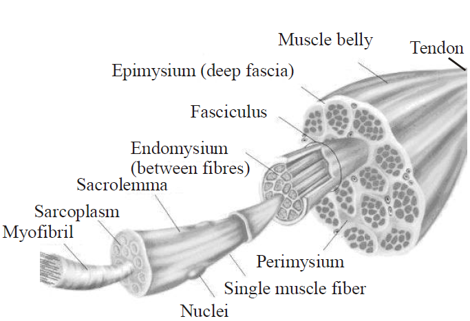
Fig. : Sectional view of Muscle
- A characteristic feature of the muscle fibre is the presence of a large number of parallelly arranged filaments in the sarcoplasm called myofilaments or myofibrils as a contractile element.
- Each myofibril has alternate dark and light bands on it.
- The light bands contain actin and is called I-band or isotropic band, whereas the dark band called ‘A’ or anisotropic band and contains myosin. Both the proteins are arranged as rod-like structures, parallel to each other and also to the longitudinal axis of the myofibrils.
- Actin filaments are thinner as compared to the myosin filaments, hence are commonly called thin and thick filaments respectively.
- In the centre of each ‘I’ band is an elastic fibre called ‘Z’ line which bisects it. The thin filaments are firmly attached to the ‘Z’ line. The thick filaments in the ‘A’ band are also held together in the middle of this band by a thin fibrous membrane called ‘M’ line.
- The ‘A’ and ‘I’ bands are arranged alternately throughout the length of the myofibrils.
- The portion of the myofibril between two successive ‘Z’ lines is considered as the functional unit of contraction and is called a sarcomere. Hence, sarcomere are the contractile units of myofibrils.
- In a resting state, the edges of thin filaments on either side of the thick filaments partially overlap on free ends of the thick filaments leaving the central part of the thick filaments. This central part of thick filament, not overlapped by thin filaments is called the ‘H’band or H-zone.
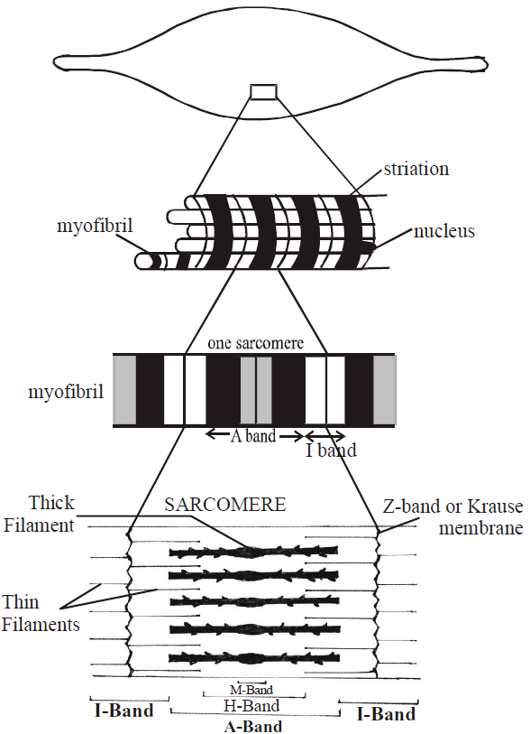
Fig. : Detailed structure of a muscle fibre
STRUCTURE OF CONTRACTILE PROTEINS
- The muscle fibres are composed of 20% protein, 75% water and remaining 5% is composed of inorganic and organic matter.
- Muscle proteins are myosin, actin, tropomyosin and troponin.
MYOSIN
- The thick filaments are made up of myosin molecule.
- Each myosin molecule contains two identical heads with pairs of helical strands which form the tail. The upper surface of each globular head contains actin binding site and ATP binding site.
ACTIN
- It is the main constituent of thin filament.
- The actin filament resembles two strings of beads twisted into a double helix. Each bead is a molecule of G-actin (globular actin) having 55Å diameter.
- It shows high affinity for calcium ions. In presence of salts and ATP, it is converted into fibrous actin (F-actin).
TROPOMYOSIN
- It is a two stranded α-helical rod which is located in the groove between the two helical strands of actin.
- A troponin complex is attached to the tropomyosin at regular interval of about 385 Å.
TROPONIN
- It is a globular protein.
- It consists of three polypeptide chains, the calcium binding subunit, the inhibitory subunit and the tropomyosin-binding subunit.
- It is an important control protein.
- It has high affinity for calcium. When calcium (Ca2+) binds with the troponin, the active sites of actin protein are exposed.

Fig. : An Actin and Myosin filaments
MECHANISM OF MUSCLE CONTRACTION
- Mechanism of muscle contraction is explained by the sliding filament theory which states that contraction of a muscle fibre takes place by the sliding of the thin filaments over the thick filaments.
- Sliding filament theory of muscle contraction was proposed by A.F. Huxley and H.E. Huxley in 1954.
- According to this theory, during muscle contraction, actin filaments slide inward on the myosin filament of A-band with the help of cross bridges to reduce the length of the sarcomere.
- Muscle contraction is initiated by a signal sent by the central nervous system (CNS) via a motor neuron.
- A neural signal reaching this neuromuscular junction or motor end plate releases a neurotransmitter (acetylcholine) which generates an action potential in the sarcolemma that spreads through the muscle fibre and causes the release of calcium ions into the sarcoplasm.
- Calcium ions trigger the muscle contraction process.
- Actin and myosin get combined in presence of ATP and Ca++ to form actomyosin to provide contraction in the muscle
Myosin + Actin  Actomyosin.
- Energy released by the oxidation of food in the form of ATP is broken down into ADP, phosphorous and energy in the presence of enzyme myosin ATPase, Ca++ and magnesium (Mg++) ions.
ATP + H2O ADP + Pi + Energy
- The energy is used up in the contraction of muscle fibre.
- When muscle contracts, creatine phosphate breaks down to produce ATP.
ADP + Creatine phosphateATP + creatine
- After that, Cori cycle takes place in liver and muscle (proposed by Coli and Coli). During glycolysis, the breakdown of glycogen takes place in the muscles and lactic acid and energy is released. Energy released is thus, used for
- the reformation of creatine phosphate from creatine.
- 4/5 of lactic acid in converted back into glycogen and remaining 1/5 is oxidized into CO2 and water.
- During muscle contraction
- Both H and I zones progressively shorten and eventually disappear.
- Length of A band remains unchanged
- Distance between two lines shorten and size of sarcomere decreases by 60 – 70%.
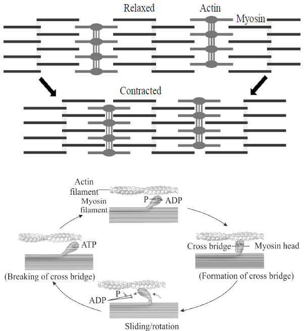
Fig. : Stages in cross bridge formation, rotation of head and breaking of cross bridge
MAJOR TYPES OF SKELETAL MUSCLE FIBRE
- Based upon the sarcoplasmic contents, situation of nuclei and number of formed elements, two types of fibres are recognised in striated muscle – white & red fibre.
- White muscle fibres are fast muscle fibre which lack myoglobin and have fewer mitochondria. These muscle fibres perform fast work for short duration as a result of which these fibres get fatigued quickly. E.g., eyeball muscle, flight muscle of sparrow.
- Red muscle fibre are slow muscle fibre possessing red haem protein (myoglobin) and abundant mitochondria. These muscle fibres can perform slow sustained work over long period without getting fatigued. E.g., extensor muscle of back and flight muscle in kites.
SKELETAL SYSTEM
- Skeletal system consists of a framework of bones and a few cartilages.
- Bone and cartilage are specialized connective tissues. The former has a very hard matrix due to calcium salts in it and the latter has slightly pliable matrix due to chondroitin salts.
- Skeletal system are commonly divided into three types – external (an exoskeleton), internal (an endoskeleton) and fluid based (a hydrostatic skeleton).
- Exoskeleton is found in both invertebrates and vertebrates (hair, nail etc.). It is hard, protective and supportive framework present outside the body.
- Endoskeleton consists of those hard parts which are present inside the body of the animal. These skeleton is found in corals, echinoderms and vertebrates.
- Hydrostatic skeletons are located internally in cnidarians, annelids and those animals which can move by contracting the muscles surrounding the fluid filled pouch, creating pressure within the pouch that cause movement.
- Human skeleton consists of 270 bones, which are fused to become 206 and out of which 6 bones occur as ear ossicles. The remaining 200 bones are divided into axial and appendicular skeleton.

Fig. : Divisions of skeletal system
AXIAL SKELETON
Axial skeleton comprises 80 bones, distributed along the main axis of the body. The skull, vertebral column, sternum and ribs constitute axial skeleton.
SKULL
- Skeleton of head is called skull. It consists of 2 main parts - cranium and face.
- Cranium bones contain sinuses. Sinuses are air spaces lined with mucous membrane that reduces weight of skull and gives resonant sound to the voice.
- Cranium (brain box) is formed of 8 bones. They form the hard protective outer covering for the brain.
- The facial region is made up of 14 skeletal elements which form the front part of the skull.
- A single U-shaped bone called hyoid is present at the base of the buccal cavity and it is also included in the skull.
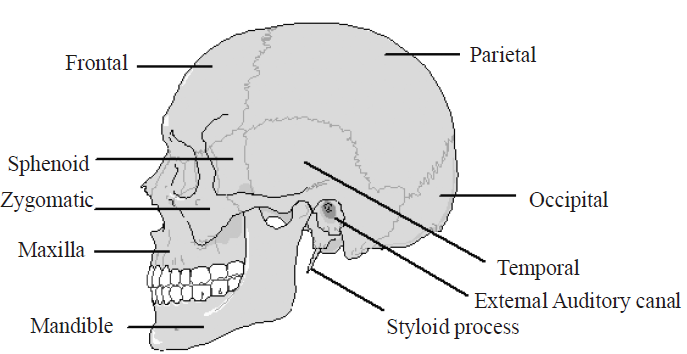
Fig. : Human skull bones
VERTEBRAL COLUMN
- Vertebral column (backbone) is a string like vertebrae which extends in the mid axis of the back (posterior) part of our trunk from head to the lower (inferior) extremity of trunk. Together with the sternum and rib, it forms the supporting framework of our trunk.
- Each vertebra has a central hollow portion (neural canal) through which the spinal cord passes.
- It supports and rotates the head, suspends the viscera, protects vital organs, provides attachment to limb girdles, facilitates some movement of the trunk and houses the spinal cord.
- Vertebral column makes two-fifth of total weight of body.
- The length of human vertebral column is 71 cm (28 inch) in adult male and about 61 cm (24 inch ) in an average adult female.
- The vertebral column is differentiated into cervical (7), thoracic (12), lumbar (5), sacral (1-fused) and coccygeal (1-fused) regions starting from the skull.
(1) Cervical vertebrae
The number of cervical vertebrae are seven in almost all mammals including human beings.
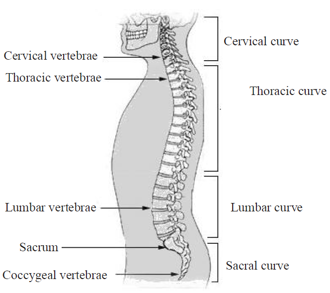
Fig. : Vertebral column
Atlas vertebra is the first cervical vertebra, whose body is formed of vertebral arch transverse process. It supports the globe of the head like the earth by the atlas (superman). Centrum and neural spine are absent. Transverse process are long with transverse foramen.
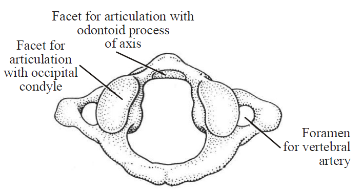
Fig. : Atlas
The second cervical vertebra is axis vertebra. Odontoid process and dens are found, which is a modified form of the centrum of atlas. Odontoid process fits into the canal of atlas to provide head with sideways rotation. Transverse process are small.
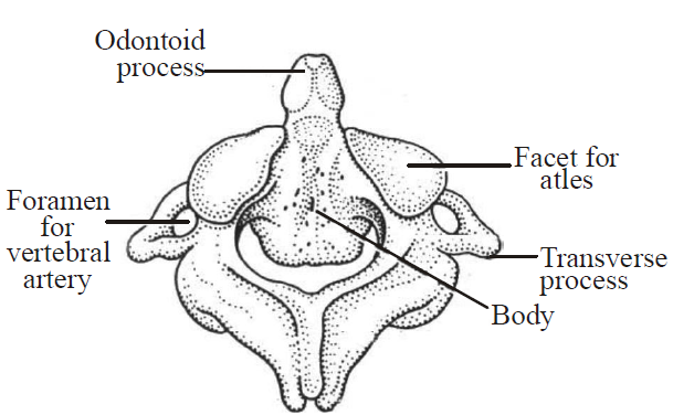
Fig. : Axis
Typical cervical vertebra contains long neural spines. Centrum is acoelous and transverse process are long. Vertebrarterial canal are found which is also called foramina transversia.
(2) Thoracic vertebra
- Centrum is acoelous and neural canal is formed by the union of two neural arches.
- Neural spine is a flat & long directed backward.
- Club shaped transverse process is present
- Neural arch is found with superior articular process.
- Two demifacets for articulation of head of a rib are present.
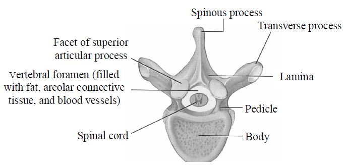
Fig. : Thoracic-vertebrae
(3) Lumbar vertebra
- Centrum is acoelus.
- Neural spine is well developed.
- Transverse process are thin and long.
- Small accessory process present near the root of each transverse process.
- It is the largest, heaviest and strongest vertebrae as they bear the weight of the abdominal viscera.
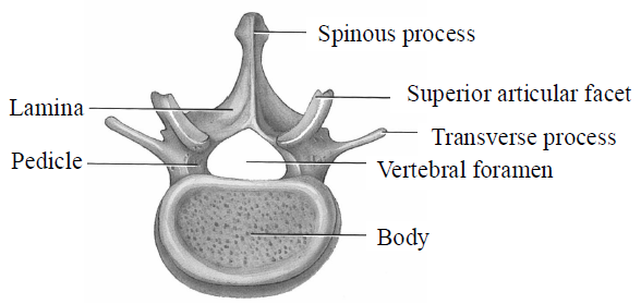
Fig.: Lumbar vertebrae
(4) Sacrum vertebra
- It is a triangular bone formed by the fusion of 5 sacral vertebra. Fusion normally begins between 16 to 18 years of age and is completed by 30 yrs. of age.
- It serves as a strong foundation for pelvic girdle.
- Sacrum with 4 pairs of anterior and posterior sacral foramina is found.
- Lateral part of sacrum articulate with ilium of hip bone.
- Female sacrum is shorter, wider and more curved between S2 and S3 whereas the male sacrum is longer, narrower, and less curved.
In birds, some of the vertebrae are fused to form synsacrum.
[Last thoracic + Lumbar + Sacral + One or two caudal]
[Last thoracic + Lumbar + Sacral + One or two caudal]
(5) Coccyx vertebra
- It is formed by the fusion of the four coccygeal vertebrae. Fusion generally occurs between 20 and 30 years of age.
- It is a small triangular bone and the last section of backbone.
- Two coccygeal cornua project up to articulate with sacral cornua.
- Transverse process are rudimentary.
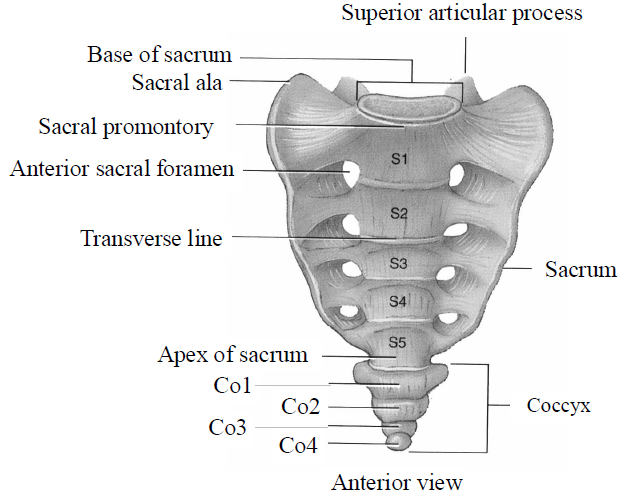
Fig.: Human sacrum and coccyx
- Human vertebral formula is C7T12L5S5Cd4.
- Function of vertebral column is to carry weight of body during motions as well as while standing and it gives flexibility to one animal during movement of head.
- Displacement of the vertebrae from its normal position due to displacement or degeneration of a part of the intervertebral disc is called slip disc.
STERNUM
- It is a flat, narrow and elongated bone of chest. It is commonly called breast bone.
- It is absent in fish, turtle and associated with pectoral girdle in amphibia.
- In man, it is made up of cervical manubrium (presternum), mesosternum and xiphoid process (metasternum).
- The sternum has two functions -
- It takes part in the formation of the protective thoracic basket.
- It plays a role in the respiratory mechanism.
RIBS
- The ribs are curved bars, which movably articulate with the thoracic vertebrae at the back and with the sternum in front. All collectively form a bony cage, the thoracic basket.
- There are 12 pairs of ribs. Each rib is a thin flat bone connected dorsally to the vertebral column and ventrally to the sternum.
- It has two articulation surfaces on its dorsal end and is hence called bicephalic.
- First seven pairs of ribs which are attached to the thoracic vertebrae and ventrally connected to the sternum with the help of hyaline cartilage are called true ribs.
- The 8th, 9th and 10th pairs of ribs do not articulate directly with the sternum but join the seventh rib with the help of hyaline cartilage. These are called vertebrochondral (false) ribs.
- Last 2 pairs (11th and 12th) of ribs are not connected ventrally and are therefore, called floating ribs.
- Thoracic vertebrae, ribs and sternum together form the rib cage.
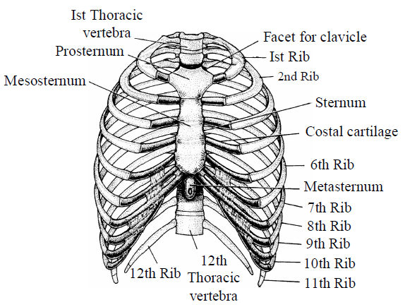
Fig. : Showing Sternum and ribs
- The ribs serve three important functions -
- They protect the heart, large blood vessels and lungs.
- They bear respiratory muscle (external and internal intercostal muscles).
- Lower two pair of ribs (11th and 12th) protect the kidney.
APPENDICULAR SKELETON
- Appendicular skeleton is made up of girdles (pectoral and pelvic) and limb bones (forelimb & hindlimb).
- Pelvic and pectoral girdle supports hindlimbs & forelimbs respectively.
- Each girdle is formed of two halves.
- The appendicular skeleton consists of 126 bones.
PECTORAL GIRDLE
- Pectoral (shoulder) girdle consists of 4 bones - 2 clavicles and 2 scapulae.
- Clavicle or Collarbone is a slender S-shaped bone that articulates with the manubrium of the sternum.
- Scapula is a large triangular flat bone situated in the dorsal part of the thorax between the second and seventh ribs. The dorsal, flat, triangular body of scapula has a slightly elevated ridge called the spine which projects as a flat, expanded process called the acromion. Below the acromion is a depression called the glenoid cavity which articulates with the head of the humerus to form the shoulder joint.
- Coracoid process is a knob like inwardly bent fused scapula blade.
PELVIC GIRDLE
- Pelvic girdle (also called hip girdle) is formed by two innominate ( = no name) bones (coxal bones).
- Each coxal bone is formed by the fusion of three bones – ilium, ischium and pubis. At the point of fusion of the above bones is a cavity called acetabulum to which the thigh bone articulates.
- Ilium is a short and straight bone, forming the upper broadest part of coxa and prominence.
- Ischium is an elongated bone, running parallel to vertebral column. It forms the medial portion of the lower part of coxa.
- Pubis is the smaller bone and form anterior portion of the lower part of the coxa of hip.
- Obturator foramen is present as a large oval gap between the pubis and ischium. The foramen forms passage for nerves and blood vessels.
- The two halves of the pelvic girdle meet ventrally to form the pubic symphysis containing fibrous cartilage.
- The pelvic girdle serves several important functions in the body. It supports the weight of the body from the vertebral column. It also protects and supports the lower organs, including the urinary bladder, the reproductive organs and the developing facts in a pregnant woman.
- The pelvic girdle differs between men and women. In man, the pelvis is more massive. In a woman, the pelvis is more delicate. These differences reflect the woman's role in pregnancy and delivery of children.
LIMB BONES
Limb are of two types - fore limb and hind limb.
(1) Bones of fore limbs
- Each fore limb consists of 30 bones.
- Humerus or arm bone or bone of the upper arm, is the longest and largest bone of upper limb.
- It articulates proximally with scapula and distally at the elbow with both ulna and radius.
- Humerus is characterized by presence of deltoid tuberosity for the attachment of muscles.
- Distal end of humerus at the elbow joint is like pulley and called trochlea. Its groove is called the olecranon fossa whose basal part is marked by a supratrochlear foramen for the passage of brachial artery and nerve.
- Humerus is characterized by arterial foramen.
- Head of the humerus articulates with the glenoid cavity of pectoral girdle.
- Radius is present towards thumb side and ulna is present towards little finger side.
- Radius is smaller and ulna is larger.
- Styloid process is present in distal end of ulna and radius both.
- Olecranon process is present in ulna. Proximally, which forms prominence of elbow.
- Trochlear notch is formed by ulna which is also known as sigmoid notch.
- Carpals or wrist bone are eight in number, joined to one another by ligaments. Carpals are arranged in 2 rows, with 4 bones in each row.
- Metacarpals are 5 in number, and phalanges are - 14.
- Phalangeal formula is 2, 3, 3, 3, 3.
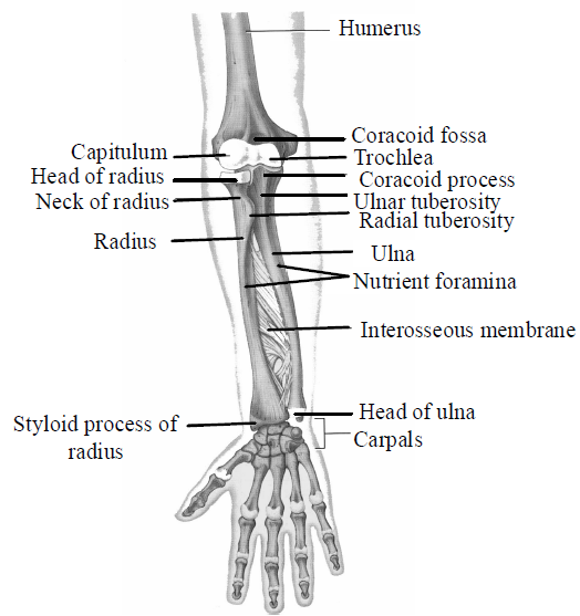
Fig. : Radio-ulna
(2) Bones of Hind Limbs
- Bones of lower limb (Hind limbs) of man are
- Thigh - Femur = 1 bone
- Knee - Knee cap - patella = 1 bone
- Leg - Tibia + Fibula = 2 bone
- Ankle - 7 tarsals = 7 bone
- Sole - 5 Metatarsals = 5 bone
- Phalangeal formula - 2, 3, 3, 3, 3 = 14 bone
- Total = 30 bone
- Bones of upper limb (Fore limbs) of man are
- Arm - Brachium - Humerus = 1 bone
- Forearm -Antibrachium - Radius + Ulna = 2 bone
- Wrist-Carpus = 4 + 4 carpals = 8 bone
- Palm - Metacarpals - 5 metacarpals = 5 bone
- Fingers - Phalangeal formula - 2, 3, 3, 3, 3 = 14 bone
- Total = 30 bone
- Femur is the longest, largest and strongest bone of the body.
- Fovea capitis is depression in head of femur.
- Femur is known as bone of thigh.
- Greater trochanter, lesser trochanter, 3rd trochanter are present in femur of thigh and buttock muscles.
- Patellar groove in found in distal end of femur.
- Patella forms knee cap.
- Patella is formed by sesamoid bone. Fabella is also example of sesamoid bone.
- Tibia is larger, also called shin bone. It bears a weight of body.
- Fibula is smaller and associated with knee joint.
- Tarsal bones are seven and metatarsals are five.
- Phalanges are fourteen. Its formula is 2, 3, 3, 3, 3
- Ankle bones have 7 tarsals and arranged in two rows.

Fig. : Femur (Anterior view)
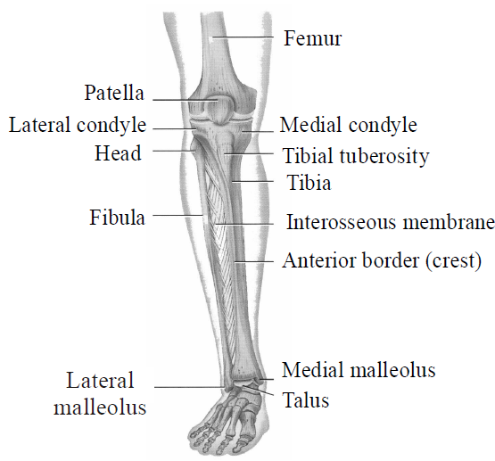
Fig. : Tibia-fibula
JOINTS
- The structural arrangement of tissue by which bones are joined together are called joints.
- Joints have been classified into three major structural forms - fibrous, cartilaginous and synovial.
FIBROUS JOINTS
Fibrous joints are immovable or fixed joints. These do not show any movement due to presence of strong and tough white cartilaginous fibres. E.g., joints in tooth sockets and between skull bones.
CARTILAGINOUS JOINTS
- Cartilaginous joints are slightly movable joints.
- In cartilaginous joints, the bones involved are joined together with the help of cartilages. The joint between the adjacent vertebrae in the vertebral column is of this pattern.
SYNOVIAL JOINTS
- Synovial joints/freely movable joints are perfect joints in which bones are not fused with each other.
- Synovial joints are characterized by the presence of a fluid filled synovial cavity between the articulating surfaces of the two bones. Such an arrangement allows considerable movement of nutrients and respiratory gases. These joints help in locomotion and many other movements.
- Synovial joints are surrounded by a tubular articular capsule. The articular capsule consists of two layers - outer fibrous capsule and inner synovial membrane.
- The synovial membrane secretes synovial fluid which lubricates and provides nourishment to articular cartilage.
- In old age, stiffness of joints is due to the decrease in synovial fluid.
- Structural arrangement of a perfect joint permits considerable movement of articulating bones without danger of friction.
- Due to the elasticity of the ligaments of the wall of joint capsule, articulating bones automatically return back to their normal positions after movements.
Synovial joints are further classified according to the movements they permit, like - Ball and socket joint (between humerus and pectoral girdle), Hinge joint (knee joint), Pivot joint (between atlas and axis and radius and ulna), Gliding joint (between the carpals), Saddle joint (between carpal and metacarpal of thumb) and ellipsoid/condyloid joint (Wrist joint and metacarpophalangeal joints)
DISORDERS OF MUSCULAR AND SKELETAL SYSTEM
- Myasthenia gravis is an autoimmune disorder. It affects neuromuscular junction leading to fatigue, weakening and paralysis of skeletal muscle.
- Muscular dystrophy is the progressive degeneration of skeletal muscle mostly due to genetic disorder.
- Tetany is a rapid spasm (wild contractions) in muscle due to low Ca++ in body fluid.
- Arthritis is a painful condition of the joints. It may be preceded or accompanied by a period of fatigue and a feeling of stiffness.
- Osteoporosis is a hereditary disease, characterized by decreased bone mass and increased chances of fractures. Decreased levels of estrogen is a common cause.
- Gout is the accumulation of uric acid crystals in the region of joints which results in painful movements.
- Sprain refers to injury to a joint capsule, typically involving a stretching or tearing of tendons or ligaments. Unfortunately, both these structures have much poorer regenerative power than bone, and once stretched, often remain weak. Sprain is often considered a minor disorder, but it may become chronic.
- Fracture is a break of a bone. Fracture occurs rarely in children. The bones of children have a large quantity of organic matter and are, therefore, very flexible and less likely to break. With advancing age, mineral matter (calcium phosphate) is deposited in the bones. This decreases the organic matter, making the bones hard and brittle. Thus, old people are more liable to fracture of bones.
Study Notes for NEET/AIIMS/JIPMER


