


CELL CYCLE AND CELL DIVISION
INTRODUCTION
- Growth and reproduction are two major characteristics of living organisms which are also shown by their individual cells. A young cell grows in size. There is replication of genetic material followed by division of the cell into two. The number of cells increases with each cycle of growth and division. A large sized cell population is produced and differentiation leads to the formation of tissues, organs and organ systems.
- Prevost and Dumas (1824) are the first to study cell division during the cleavage of zygote of frog.
- Rudolf Virchow (1855) observed that new cells always develop from pre-existing cells. He also gave cell lineage theory or law of cell lineage and doctrine of genetic continuity.
- Cell division is the process by which a mature cell divides and forms two nearly equal daughter cells which resemble the parental cell in a number of characters.
- The cell which undergoes division is called mother cell or parent cell. The newly formed cells are known as daughter cells.
- In unicellular organisms, cell division is the means of reproduction by which the mother cell produces two or more new cells. In multicellular organisms also, new individual develops from a single cell.
CELL CYCLE
- Cell division is a biological process in all living organisms.
- Although cell growth (in terms of cytoplasmic increase) is a continuous process, DNA synthesis occurs only during one specific stage in the cell cycle. The replicated chromosomes (DNA) are then distributed to daughter nuclei by a complex series of events during cell division. These events are themselves under genetic control.
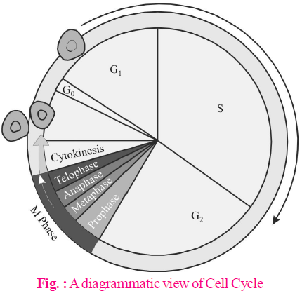
- The sequence of events which occur during cell growth and cell division are collectively called cell cycle.
PHASES OF CELL CYCLE
The period required to complete one cell cycle (from beginning of one cell division to the beginning of next) is called generation time. It is 24 hours in human cells and 90 minutes in yeast. Cell cycle is simpler in prokaryotes and more complex in eukaryotes.
The cell cycle is divided into two basic phases :
- Interphase
- M Phase (Mitosis phase)/Dividing phase
INTERPHASE
- It is the period between the end of one cell division to the beginning of the next cell division.
- It is a highly metabolically active phase in which cell prepares itself for the next cell division.
- All mature cells of the body, therefore, occur in interphase. Nerve cells of mammals have the longest interphase and thus, do not divide after their formation at birth.
Interphase is completed into three successive stages.
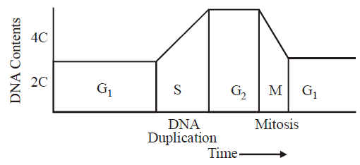
(1) G1 phase/Post mitotic/Pre-DNA synthetic phase/gap-I
G1 phase corresponds to the interval between mitosis and initiation of DNA replication.
Following events take place during this phase
- Intensive cellular synthesis.
- Synthesis of rRNA, mRNA ribosomes and proteins.
- Metabolic rate is high.
- Cell size increases.
- Synthesis of enzymes, amino acids, nucleotides etc. but there is no change in DNA amount.
(2) S-phase/Synthetic phase
S or synthesis phase marks the period during which DNA synthesis or replication takes place.
Following events take place during this phase
- DNA replicates and its amount becomes double. If the initial amount of DNA is denoted as 2C then it increases to 4C.
- Synthesis of histone proteins and NHC (non-histone chromosomal proteins).
- Duplication of centrioles in the cytoplasm.
(3) G2-phase/Pre mitotic/Post synthetic phase/Gap-II
Following events take place during this phase
- Mitotic spindle protein (tubulin) synthesis begins.
- Chromosome condensation factor appears.
- Synthesis of 3 types of RNA, NHC proteins, and ATP molecule.
- Repair of damaged DNA occurs.
- The cells that do not divide further exit G1 phase to enter an inactive stage called quiescent stage (G0) of the cell cycle. Cells in this stage remain metabolically active but no longer proliferate unless called on to do so depending on the requirement of the organism.
M -PHASE/DIVIDING PHASE/MITOTIC PHASE
- It is the phase of actual cell division.
- It is divided into two phases - karyokinesis (division of nucleus) and cytokinesis (division of the cytoplasm).
- Time period for G1, S, G2 and M-phase is species specific under specific environmental conditions e.g., 20 minutes for bacterial cell, 8-10 hours for intestinal epithelial cells, onion root tip cells may take 20 hours. 24 hours for human cells and 90 minutes for yeast cells to complete these phases of cell cycle.
- It is of three types – amitosis, mitosis and meiosis.
AMITOSIS
- Amitosis is also called as direct cell division.
- In this division, there is no differentiation of chromosomes and spindle. The nuclear envelope does not degenerate. The nucleus elongates and constricts in the middle to form two daughter nuclei. This is followed by a centripetal constriction of the cytoplasm to form two daughter cells.
- Examples : Prokaryotes, protozoans, yeasts, foetal membrane of mammals, cartilage of mammals etc.
MITOSIS
- Mitosis is also called indirect cell division or somatic cell division or equational division.
- In this, mature somatic cells divide in such a way that chromosome number is kept constant that is, number of chromosomes in daughter cells equal to those in the parent cell.
- The growing regions of plants have meristematic cells (e.g. these cells are found in apical portion of root and stem and in the expanding leaf) in which mitosis takes place.
KARYOKINESIS
It comprises of four phases i.e., Prophase, Metaphase, Anaphase, Telophase.
(1) Prophase
It is the longest phase of karyokinesis.
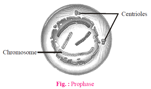
- Chromatin fibres thicken and shorten to form chromosomes which may overlap each other and appear like a ball of wool.
- Each chromosome divides longitudinally into 2 chromatids which remain attached to centromere.
- Nuclear membrane starts disintegrating except in dinoflagellates.
- Nucleolus starts disintegrating.
- Spindle formation begins.
- In cytoskeleton, golgi complex, ER, etc. disappear.
- In animal cells, centrioles move towards opposite sides.
(2) Metaphase
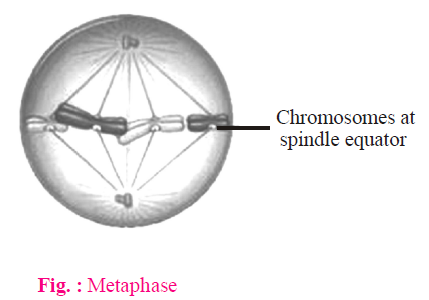
- Chromosomes become maximally distinct i.e., size can be measured.
- Chromosomes move towards equatorial plane of spindles called congression and become arranged with their arms directed towards pole and centromere towards equator.
- Spindle fibres attach to kinetochores.
- Metaphase is the best stage for studying chromosome morphology (structure, size, number).
(3) Anaphase
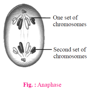
- Centromere splits from the middle and two chromatids get separated.
- Both the chromatids move towards opposite poles due to repulsive force called anaphasic movement.
- Anaphasic movement is brought about by the repolymerisation of continuous fibres and depoly-merisation of chromosomal fibres. Formation and expansion of interzonal fibres occur.
- The centromere faces towards equator.
- Shape of chromosome is best studied at anaphase.
(4) Telophase
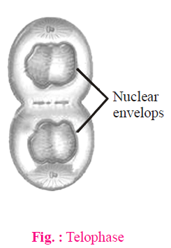
- Chromosomes reach the poles by the spindle fibers and form two groups.
- Chromosomes begin to uncoil and form a chromatin net.
- The nuclear membrane and nucleolus reappear.
- In animal cells, astral rays and spindle fibres completely disappear in telophase. The two centriole pairs organise themselves into centrosomes. In plant cells, the spindle fibres disappear from near the poles but remain intact towards the equator.
- Golgi complex and ER etc., reform.
CYTOKINESIS
- Cytokinesis is the division of cell having undergone karyokinesis to produce two daughter cells each with a daughter nucleus. It begins in mid-anaphase and is generally completed along with the completion of telophase.
- Cytokinesis is by 2 methods:
- Cell furrow method : This is characteristic of animal cells.
A dense vesicular and fibrous structure is formed in the equatorial region of the spindle simultaneously with its depolymerisation. It is called mid body.
A circular constriction or invagination appears at the centre or equator and forms a furrow. The constriction or furrow deepens centripetally and divides the mother cell into two daughter cells.
- Cell plate method : This is characteristic of plant cells. Here, vesicles provided by Golgi apparatus unite to form phragmoplasts, which join to form the cell plate. Cell plate is first laid down in the centre and then proceeds towards periphery (i.e., centrifugal plate-formation). Cell wall materials are now laid down on both sides of cell plate thus, forming two daughter cells.
TYPES OF MITOSIS
- Intranuclear or Promitosis : Nuclear membrane is not disintegrated and spindle is formed inside the nuclear membrane e.g., Protozoans (Amoeba) and yeast. It is so as centriole is present within the nucleus.
- Extranuclear or Eumitosis : In this, nuclear membrane is disintegrated and spindle is formed outside nuclear membrane e.g., in plants and animals.
- Endomitosis : Chromosomes and their DNA duplicate but fail to separate which leads to polyploidy e.g., in liver of man, both diploid (2N) and polyploid cells (4N) have been reported. It is also called endoduplication and endopolyploidy.
- Dinomitosis : In this, nuclear envelope persists but microtubular spindle is not formed. During movement, the chromosomes are attached with nuclear membrane, e.g., dinoflagellates.
SIGNIFICANCE OF MITOSIS
- It keeps the chromosome number constant and maintains genetic stability in daughter cells. All the cells have similar genetic constituents.
- It provides new cells for repair and regeneration and for healing of the wounds. (e.g., cells of the upper layer of the epidermis, cells of the lining of the gut, and blood cells).
- It helps in asexual reproduction by fragmentation, budding, stem cutting, etc.
- Somatic variations when maintained by vegetative propagation can play an important role in speciation.
MITOGENS AND MITOTIC POISONS
The agents which stimulate cell division are called mitogens, e.g., cytokinins, some steroids, however, some chemicals inhibit cell division, these are called mitotic poisons, e.g., azides, cyanides, colchicine, chalones, etc. Colchicine is an alkaloid derived from Colchicum autumnale. It interferes with spindle formation and arrests cell division at metaphase. Polyploidy in plants can be induced by the application of colchicine.
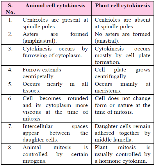
Table : Difference between the Cytokinesis of Animal cell and Plant cell.
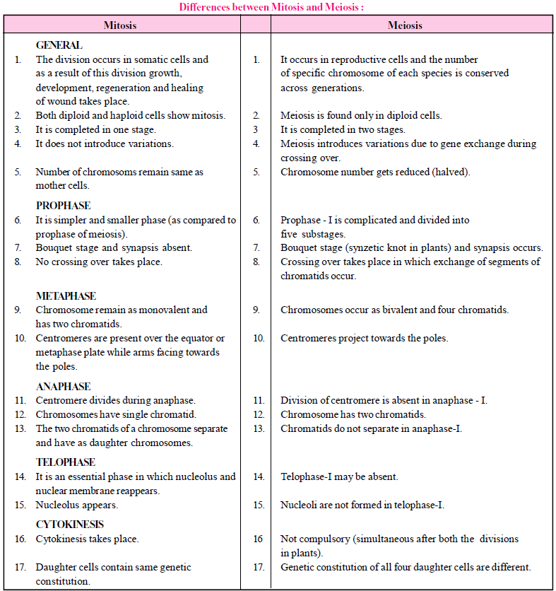
MEIOSIS
- It is a division that occurs in mature diploid reproductive cells (2x) in which the nucleus divides twice but chromosome (DNA) replicates only once to form four haploid cells, each having half the number of chromosomes present in the parent cell. As it causes a reduction in the number of chromosomes, it is known as reduction division. Meiosis in a cell occurs only once. The so formed haploid cells do not further undergo meiosis because there is no synaptonemal complex in haploid genome.
- It is reported in diploid germ cells of sex organs (e.g., primary spermatocytes of testes to form male gametes called spermotozoa and primary oocytes to form female gametes called ova in animals) and in pollen mother cells (microsporocytes) of anther and megasporocyte of ovules of ovary of flowers in plant to form the haploid spores. The study of meiosis in plants can be done in young flower buds.
- Meiosis I is initiated after the parental chromosomes have replicated to produce identical sister chromatids during the S phase.
- Meiosis involves pairing of homologous chromosomes and recombination between them.
- Four haploid cells are formed at the end of meiosis II.
- Process of meiosis : Meiosis is completed in two steps, Meiosis I and Meiosis II. Each of them is further divisible into different stages as in mitosis.
Meiosis I Meiosis II
Prophase I Prophase II
Metaphase I Metaphase II
Anaphase I Anaphase II
Telophase I Telophase II
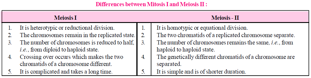
MEIOSIS-I
It results in the formation of two haploid cells from one diploid cell. The daughter cells are, therefore, haploid but with 2n DNA content. It is divided into four phases i.e., prophase, metaphase, anaphase, telophase.
(1) Prophase-I
It is the longest phase of karyokinesis of meiosis. It is again divisible into five subphases i.e., leptotene, zygotene, pachytene, diplotene and diakinesis.
(a) Leptotene/Leptonema
- Chromosomes are long thread like with chromomeres (i.e. linear series of darkly stained swollen areas) on it.
- Each chromosome has its similar structure or homologue known as homologous chromosomes derived from different parents either paternal or maternal.
- Leptonema chromosomes have a definite polarization and form loops whose ends are attached to the nuclear envelope at points near the centrioles, contained within an aster. Such peculiar arrangement is termed as bouquet stage (in animals) and synzetic knot (in plants like Lilium).
- Lampbrush chromosome found in oocyte of amphibians is seen in leptotene.
(b) Zygotene/Zygonema
- Pairing or "Synapsis" of homologous chromosomes takes place in this stage.
- Paired chromosomes are called bivalents which by further molecular packing and spiralization become shorter and thicker.
- Pairing of homologous chromosomes in a zipper-like fashion.
- On observing under an electron microscope, a filamentous ladder like nucleoproteinous complex, called synaptonemal complex helps in adherence of the visible sister chromatids.
(c) Pachytene/Pachynema
- In the tetrad, two similar chromatids of the same chromosome are called sister chromatids while chromatids belonging to different chromosomes of the homologous pairs are termed as non-sister chromatids.
- Crossing over i.e., exchange of segments between non-sister chromatids of homologous chromosomes occurs at this stage.
It takes place by breakage and reunion of chromatid segments. Breakage called nicking is assisted by an enzyme endonuclease and reunion termed annealing is done by an enzyme ligase.
- A tetrad consists of two sets of homologous chromosomes each with two chromatids. Chromatids of pachytene chromosomes are attached with centromere. Each tetrad has four kinetochores (two sisters and two homologous).
- A number of electron dense bodies about 100 nm in diameter are seen at irregular intervals within the centre of the synaptonemal complex known as recombination nodules. These are believed to be sites having multienzyme recombinase complex required for crossing over.
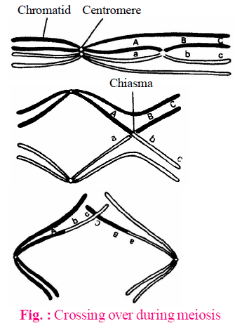
(d) Diplotene/Diplonema
- At this stage, the paired chromosomes begin to separate (desynapsis) and terminalisation starts.
- Cross is formed at the place of crossing over between non-sister chromatids.
- Homologous chromosomes move apart and remain attached to one another at specific points called chiasmata.
- At least one chiasma is formed in each bivalent.
- This stage remains as such for a long time. For example, all the oocytes of human female reach the diplotene stage in the fifth month of foetus and remain so for many years till ovulation is to occur. In oocytes of many fishes, amphibians, reptiles and birds, the bivalents elongate and become converted into lampbrush chromosomes for synthesis of specific biochemicals.
(e) Diakinesis
- Terminalization of chiasmata takes place.
- Nuclear membrane and nucleolus degenerates.
- Chromosomes recondense and tetrad moves to the metaphase plate.
- Formation of spindle occurs.
When the diakinesis of prophase-I is completed then cell enters into metaphase-I.
(2) Metaphase-I
- Chromosomes align at the equator.
- Bivalents arrange themselves in two parallel equatorial or metaphasic plates. Each equatorial plate has one genome.
- Each homologous chromosome has two kinetochores and both the kinetochores of a chromosome are joined to the chromosomal or tractile fibre of same side.
(3) Anaphase-I
- It involves separation of homologous chromosomes which start moving towards opposite poles so that each tetrad is divided into two daughter dyads. So, anaphase-I involves the reduction of chromosome number, this is called disjunction.
- Segregation of Mendelian factors or independent assortment of chromosomes takes place, in which the paternal and maternal chromosomes of each homologous pair segregate during anaphase-I thus, introducing genetic variability.
(4) Telophase-I
- Two daughter nuclei are formed but the chromosome number is half the chromosome number of mother cell.
- Nuclear membrane reappears.
- After telophase I, cytokinesis may or may not occur.
Interkinesis (Intrameiotic interphase)
Generally there is no interphase between meiosis-I and meiosis-II. A brief interphase called interkinesis, or intrameiotic interphase occurs. There is no replication of chromosomes because chromosomes are already in replicated state. Centrosome may replicate in animal cells.
Significance of Meiosis-I
- It separates the homologous chromosomes to reduce the chromosome number to the haploid state, a necessity for sexual reproduction.
- It introduces variation by forming new gene combinations through crossing over and random assortment of paternal and maternal chromosomes.
MEIOSIS-II
- It is also called equational or homotypical division because the number of chromosomes remain same as after meiosis-I.
- It involves the separation of two chromatids of each chromosome and their movement to separate cells.
- It is divided into four phases i.e., Prophase-II, Metaphase-II. Anaphase-II and Telophase-II.
(1) Prophase-II
- In contrast to meiosis I, meiosis II resembles a normal mitosis.
- The nuclear membrane disappears by the end of prophase II.
- The chromosomes again become compact.
(2) Metaphase-II
At this stage, the chromosomes align at the equator and microtubules from opposite poles of the spindle get attached to kinetochores of sister chromatids.
(3) Anaphase-II
It begins with the simultaneous splitting of the centromere of each chromosome (which was holding the sister chromatids together), allowing them to move towards opposite poles of the cell.
(4) Telophase-II
- Meiosis ends with telophase II, in which chromosomes once again get enclosed by a nuclear envelope; cytokinesis follows resulting in the formation of tetrad of cells i.e., four haploid daughter cells.
- Cytokinesis-II is always present and occurs by cell furrow formation in animal cells and cell plate formation in plant cells.
Types of meiosis II
- Gametic/Terminal meiosis : In many protozoans, all animals and some lower plants, meiosis takes place before fertilization during the formation of gametes.
- Zygotic or Initial meiosis : In fungi, certain protozoan groups, and some algae, fertilization is immediately followed by meiosis in the zygote, and the resulting adult organisms are haploid. Such a meiosis is said to be zygotic or initial. This type of life cycle with haploid adult and zygotic meiosis is termed the haplontic cycle.
- Sporogenetic / Intermediate meiosis : Spore mother cells of sporophytic plant undergo meiosis to form haploid spores in the sporangia. Haploid spore germinates to form haploid gametophyte which produces haploid gametes by mitosis. Haploid gametes fuse to form a diploid zygote which develops into diploid sporophyte by mitotic divisions. e.g., in higher plants like pteridophytes, gymnosperms and angiosperms.
Significance of Meiosis-II
- Constancy of chromosome number in successive generations is brought by this process.
- It helps in introducing variations and mutations.
- It maintains the amount of genetic material.
- The four daughter cells will have different types of chromatids.
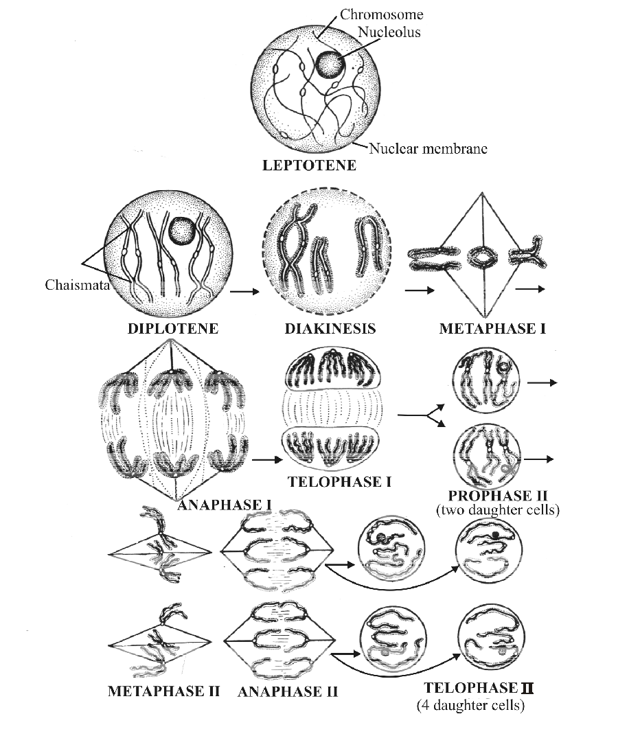
Fig. : Stages in Meiosis
Study Notes for NEET/AIIMS/JIPMER


