


BIOLOGICAL CLASSIFICATION
INTRODUCTION
- Identification of differences among organisms and placing them into groups that reflect their most significant features and relationship is called biological classification.
- The purpose of biological classification is to organise the vast number of known plants into categories that could be studied easily.
- Organisms have been classified from different points of view at different times.
- Biological classification was first proposed by Aristotle who used simple morphological characters to classify plants and animals.
ARTIFICIAL CLASSIFICATION
In this type of classification, plants are classified on the basis of one or two morphological characters i.e. overall morphology is not considered.
- Artificial system of classification was adopted by Pliny the Elder for animals on the basis of habitat, e.g. land, air and water.
- Classification proposed by Linnaeus is artificial.
- Linnaeus classified plant kingdom into 24 classes on the basis of only two characters-stamens and style in his book ‘Genera Planatarium’.
NATURAL CLASSIFICATION
- In this type, plants are classified on the basis of their complete morphology. In it, the classification of whole plant is included (like stem, root, leaves, flowers etc). Maximum characters are taken as base in this classification.
- The first natural system of plant classification was proposed by Schimper (1879) followed by Eichler (1833).
- Natural classification is believed to be the best classification because it represents the natural similarities and dissimilarities of plants i.e. it represents the inter-relationship among plants.
- In this classification, the plants belonging to the same group show many similarities, while in artificial classification, the plants belonging to the same group show only, one or two similar characters. They have many dissimilarities.
- Natural classification is of two types - natural formal and natural phylogenetic.
- In Natural formal classification, the phylogeny of the plant is not considered i.e. only the morphology of the plant is considered.
- In Natural phylogenetic classification, both morphology and phylogeny are considered. In phylogenetic classification, the plants are arranged on the basis of their evolution.
- Lamarck : Proposed the term "Phylogeny".
- Ernest Haeckel : Gave the concept of phylogeny.
- Charles Darwin : Gave broad explanation of phylogeny in his book.
- Genealogy → Sequence of evolution
- Genealogy of plant kingdom : Thallophyta → Bryophyta → Pteridophyta → Gymnosperm → Angiosperm (Most advanced plants)
ADANSONIAN SYSTEM OR PHENETIC CLASSIFICATION OR NUMERICAL CLASSIFICATION
- It is proposed by "Sokel and Sneath". Plants are classified on the basis of numbers of similarities and dissimilarities.
- In this, importance to single character is not given, all characters have the same importance. While, in natural classification, floral (reproductive) characters have more importance than vegetative (root, stem and leaves) characters.
HISTORY OF TAXONOMY
ARISTOTLE & THEOPHRASTUS (370 - 285 B.C.)
Aristotle : He is the father of biology and zoology.
Theophrastus : He is known as the father of ancient plant taxonomy and father of botany.
- Both Theophrastus & Aristotle are Greek political philosophers.
- Theophrastus wrote many books on plants. Few of them are as follows :
- Historia plantarum
- Causes of plants
- Enquiry into plants
- Theophrastus gave names and descriptions of 480 plants in his book Historia plantarum.
- Theophrastus proposes the first classification of plant kingdom. He classified plant kingdom into four groups on the basis of growth habit like trees, shrubs, under shrubs, herbs. It is an artificial classification. He proposed the term annual, biennial and perennials.
CAROLUS LINNAEUS (1707 - 1778)
- He is known as the father of taxonomy, father of plant taxonomy and father of animal taxonomy.
- Linnaeus gave the two kingdom system of classification. He grouped plants and animals into kingdom plantae and kingdom animalia respectively. Linnaeus wrote many books. Some important books are:
- Hortus uplandicus - First book
- Flora lapponica
- Philosophia botanica
- Critica botanica
- Systema naturae (1737)
- Genera plantarum
- Species plantarum - last book (1753)
- In "Philosophia botanica," Linnaeus gave the principles of nomenclature.
- In "Systema naturae," Linnaeus gave the scientific names of animals. In this book, he gave the detailed description of the animal kingdom. He also gave the outline classification of plant kingdom in this book.
- In "Genera plantarum," Linnaeus gave the detailed description of plant kingdom.
- The main basis of Linnaeus classification was the "sex organs". Therefore, this classification is also known as "Sexual classification".
- In "Species plantarum," he gave the scientific names of plants. (He gave the description of 6000 plant species).
A.P. DE CANDOLLE
- He wrote the book - "Theories Elementaire de la botanique".
- He was the first to propose the significance of vascular tissue in taxonomy. On this basis of vascular tissue, he classified plants into two groups –
- Cellular plants (Non - vascular plants) - This group includes thallophyta and bryophyta.
- Vascular plants - This group includes pteridophyta, gymnosperms and angiosperms.
GEORGE BENTHAM (1800 -1884) AND JOSEPH DALTON HOOKER (1817 -1911)
They wrote the book "Genera plantarum" (1862 - 1883). In this book, Bentham and Hooker gave the biggest and natural classification of spermatophyta i.e. plants with seeds. In Genera plantarum, there is description of 202 families.
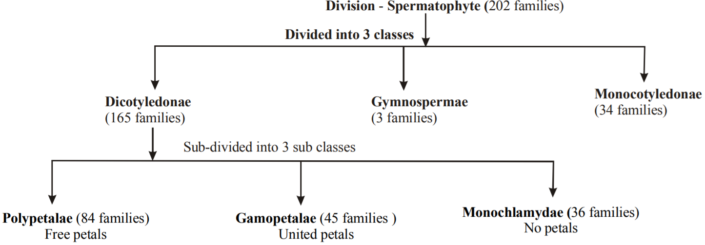
Flow chart 2.1 : Classification of spermatophyta by Bentham and Hooker
Merits of Bentham and Hooker classification
- The classification of Bentham and Hooker was natural formal.
- The classification of Bentham and Hooker was mainly based on the floral characters. This was very appreciable because floral characters are more stable than vegetative characters.
- It is the simplest classification. Therefore, the arrangement of all plants in the botanical gardens and herbarium of the world is based on it. Although it is not the best classification but yet the arrangement of plants in botanical gardens and herbarium is based on it, because it is the simpler one. The main reason for its simplicity is that this classification is based on actual observations.
Demerits of Bentham and Hooker
In this classification, the phylogeny of plants is not considered because in it gymnosperms are placed in between dicots and monocots. The sequence of evolution is as follows –
Phylogeny = Gymnosperm → Dicots → Monocots
A. W. EICHLER
- Eichler gave the first phylogenetic classification of plant kingdom.
- Eichler classified plant kingdom into two sub-kingdoms- cryptogamia and phanerogamia
- The classification of Eichler is very little phylogenetic.
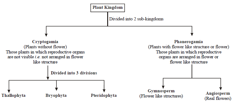
Flow chart 2.2 : Classification of plant kingdom (by Eichler)
- In this way, Eichler classified plant kingdom into five divisions and arranged them in the order of evolution (Phylogeny).
Thallophyta → Bryophyta → Pteridophyta → Gymnosperm → Angiosperm
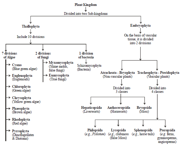
ENGLER (1844 - 1930) AND PRANTL (1849 - 1893)
- Book - "Die Naturlichen Pflanzen Familien" is written by Engler & Prantl.
- He gave the phylogenetic classification of plant kingdom. This classification was more phylogenetic as compared to Eichler's classification.
Flow chart 2.3 : Plant classification by Engler and Prantl
OSWALD TIPPO
He proposed the biggest phylogenetic classification of plant kingdom.
This classification is the complete classification of plant kingdom (Refer flowchart 2.4).
Flow chart 2.4 : Classification of Plant Kingdom by Oswald Tippo
HAECKEL
- Haeckel gave the three kingdoms (Protista, Plantae, Animalia) system of classification (1866).
- Haeckel established the kingdom Protista.
- The term 'Protista' was given by C. Cuvier.
- Haeckel grouped those living organisms in Protista which did not have tissues.
- Kingdom Protista includes prokaryotes, protozoa, porifera, algae and fungi.
- This system of classification was not accepted because it includes both prokaryotic & eukaryotic chlorophyllous and non chlorophyllous organisms together.
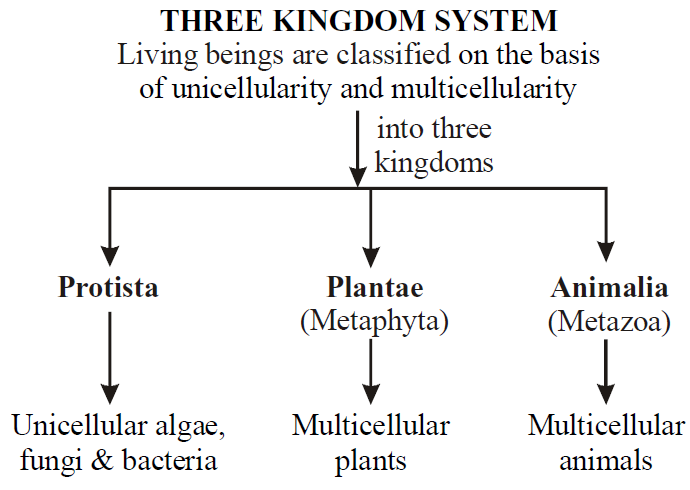
Flow chart 2.5 : Three kingdom system of classification by Haeckel
COPELAND (1956)
He gave the four kingdom system of classification.
- Mycota : Dougherty and Allen gave the name "Monera" to Mycota of Copeland. All the prokaryotes are grouped in Monera. E.g., bacteria, mycoplasma, blue green algae.
- Protista or Prototista : Copeland grouped those eukaryotes in protista, which are visually different than normal plants and animals. Eg, brown algae, red algae, fungi, protozoa
- Plantae or Metaphyta : Remaining all eukaryotic plants are grouped into this kingdom.
- Animalia or Metazoa : Remaining all eukaryotic animals are grouped into this kingdom.
R.H. WHITTAKER (1969)
- He gave the five kingdom system of classification.
- The five kingdom classification of Whittaker was based on 3 main characters –
- Complexity of cell : Cell is prokaryote or eukaryote , on this basis, Kingdom Monera is formed and all the prokaryotes are grouped into it.
- Complexity of organism : Organism is unicellular or multicellular. On this basis Kingdom Protista was formed, and all the unicellular eukaryotes were grouped into it.
- Nutrition : Organism is autotrophic or heterotrophic. On this basis Kingdom Mycota, Plantae and Animalia were formed. Except fungi (heterotrophic), all the plants are autotrophs. Therefore, fungi is separated from plants and placed in Kingdom Mycota. And remaining all the autotrophic plants are placed in Kingdom Plantae. Since all the animals are heterotrophs, therefore they are placed in the fifth kingdom i.e. kingdom Animalia.
THE FIVE KINGDOMS
- Monera : It includes all the prokaryotes (Eubacteria, Actinomycetes, blue green algae, Mycoplasma) and Akaryote (virus).
- Protista : It includes all the unicellular eukaryotes (Protozoans, Dinoflagellates, Diatoms, Euglenoids, Slime molds).
- Mycota : It includes true fungi.
- Plantae : It includes multicellular eukaryotic plants (Algae, Bryophyte, Pteridophyte, Gymnosperm and Angiosperm).
- Animalia : It includes multicellular animals.
Characteristics of five kingdoms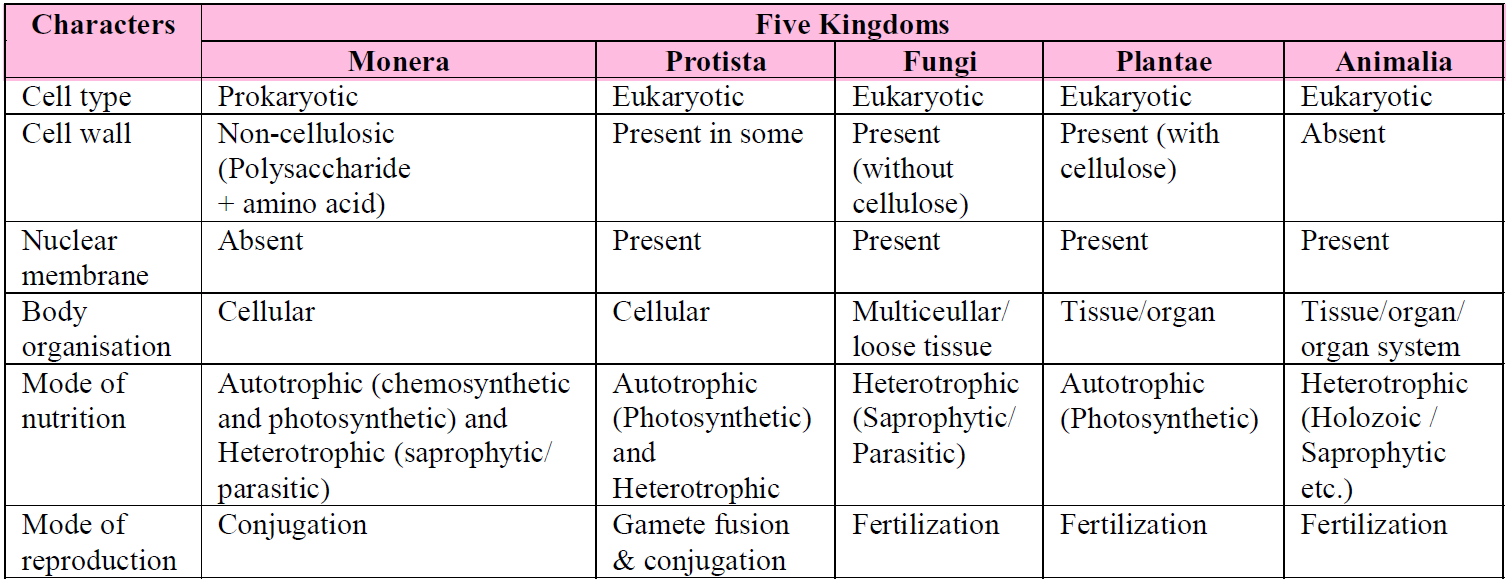
KINGDOM MONERA
- Monera (Monos - single) includes prokaryotes.
- They are typically unicellular organisms (but one group is mycelial).
- The genetic material is naked circular DNA, not enclosed by a nuclear envelope.
- Ribosomes and simple chromatophores are the only subcellular organelles in the cytoplasm. The ribosomes are 70S. Mitochondria, plastids, golgi apparatus, lysosomes, endoplasmic reticulum, centrosome, etc. are lacking.
- Sap vacuoles do not occur. Instead, gas vacuole may be present.
- The predominant mode of nutrition is absorptive but some groups are photosynthetic (holophytic) and chemosynthetic.
- The organisms are non-motile or move by beating of simple flagella or by gliding.
- Flagella, if present, are composed of many intertwined chains of a protein flagellin. They are not enclosed by any membrane and grow at the tip.
- Moneran cells are microscopic (1 to few microns in length).
- Most organisms bear a rigid cell wall (peptidoglycan).
- Reproduction is primarily asexual by binary fission or budding. Mitotic apparatus is not formed during cell division.
- It includes bacteria, actinomycetes, mycoplasma and cyanobacteria.
BACTERIA
- Bacteria are the smallest free living organisms which are mostly unicellular.
- Bacteria was discovered by Leeuwenhoek in pond water and in tartar scraped from teeth.
- Compared to many other organisms, bacteria as a group shows the most extensive metabolic diversity. Some of the bacteria are autotrophic, i.e., they synthesize their own food from inorganic substrates. They may be photosynthetic autotrophic or chemosynthetic autotrophic. The vast majority of bacteria are heterotrophs, i.e., they do not synthesise their own food but depend on other organisms or on dead organic matter for food.
- Bacteria are grouped under four categories based on their shape: the spherical Coccus, the rod-shaped Bacillus, the comma-shaped Vibrio and the spiral Spirillum.
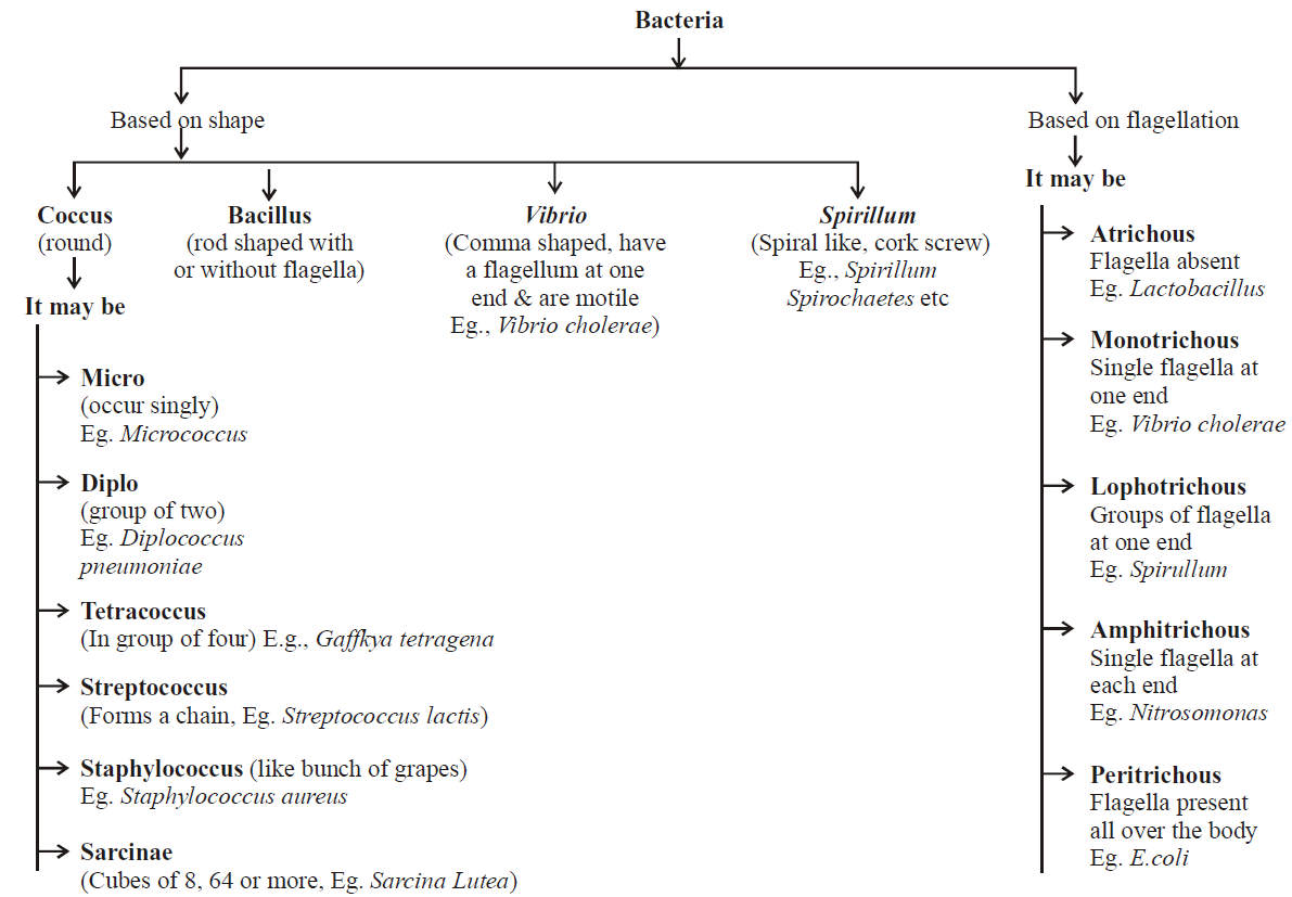
Flow chart 2.6 : Types of bacteria based on shape & flagellation
STRUCTURE OF BACTERIAL CELL
- CAPSULE : In a large number of bacteria, a slimy capsule is present outside the cell wall. It is composed of polysaccharides and the nitrogenous substances (amino acids) are also present in addition. This slime layer becomes thick called capsule. The bacteria, which form a capsule, are called capsulated or virulent bacteria. The capsule is usually found in parasitic forms, e.g., Bacillus anthracis, Diplococcus pneumoniae, Mycobacterium tuberculosis.
- CELL WALL : All bacterial cells are covered by a strong, rigid cell wall. Therefore, they are classified under plants. Inner to the capsule, cell wall is present. It is made up of polysaccharides, proteins and lipids.
In the cell wall of bacteria, there are two important sugar derivatives i.e., NAG and NAM (N-acetylglucosamine and N-acetyl muramic acid) and besides L or D-alanine,
D-glutamic acid and diaminopimelic acid are also found.
D-glutamic acid and diaminopimelic acid are also found.
- PLASMA MEMBRANE : Each bacterial cell has plasma membrane situated just internal to the cell wall. It is a thin, elastic and differentially or selectively permeable membrane. It is composed of large amounts of phospholipids, proteins and some amounts of polysaccharides but lacks sterols. It is characterized by possessing respiratory enzymes.
- CYTOPLASM : The cytoplasm is a complex aqueous fluid or semi fluid ground substance (matrix) consisting of carbohydrates, soluble proteins, enzymes, coenzymes, vitamins, lipids, mineral salts and nucleic acids. The organic matter is in the colloidal state.
The cytoplasm is granular due to the presence of a large number of ribosomes. Ribosomes in bacteria are found in the form of polyribosome. Membranous organelles such as mitochondria, endoplasmic reticulum, golgi bodies, lysosomes and vacuoles are absent. In some photosynthetic bacteria, the plasma membrane gives rise to large vesicular thylakoids which are rich in bacteriochlorophylls and proteins.
- NUCLEOID : It is also known as genophore, naked nucleus, incipient nucleus. There is nuclear material DNA which is double helical and circular. It is surrounded by some typical protein (polyamine) but not histone proteins. Histones (basic proteins) are altogether absent in bacteria.
- PLASMID : In addition to the normal DNA chromosomes, many bacteria (e.g., E.coli) have extra chromosomal genetic elements or DNA. These elements are called plasmids. Plasmids are small circular double stranded DNA molecules. The plasmid DNA replicates independently maintaining independent identity and may carry some important genes. Plasmid term was given by Lederberg (1952). Some plasmids are integrating into the bacterial DNA chromosome called episomes.
There are 3 types of plasmids :
- F-factor or fertility factor : It is responsible for transfer of genetic material.
- R-factor or resistance factor : It provides resistance against drugs.
- Colicinogenic factor : It produces colicines which kill other bacteria.
- FLAGELLA : These are fine, thread-like, protoplasmic appendages which extend through the cell wall and the slime layer of the flagellated bacterial cells. These help bacteria to swim about in the liquid medium.
Bacterial flagella are the most primitive of all motile organs. Each is composed of a single thin fibril as against the 9+2 fibrillar structure of eukaryotic cells. The flagellum is composed entirely of flagellin protein.
- PILI OR FIMBRIAE : Besides flagella, some tiny or small hair-like outgrowths are present on bacterial cell surface. These are called pili and are made up of pilin protein. They measure about 0.5 – 2 μm in length and 3 – 5μm in diameter. Fimbriae take part in attachment like holding the bacteria to solid surfaces.
Some sex pili act as conjugation canals through which DNA of one cell passes into the other cell.
STAINING OF BACTERIA
- SIMPLE STAINING : The coloration of bacteria by applying a single solution of stain to a fixed smear is termed simple staining. The cells usually stain uniformly.
- GRAM STAINING : This technique was introduced by Hans Christian Gram in 1884. It is a specific technique which is used to classify bacteria into two groups Gram +ve and Gram –ve. The bacteria are stained with weakly alkaline solution of crystal violet. The stained slide of bacteria is then treated with 0.5 percent iodine solution. This is followed by washing with water or acetone or 95% ethyl alcohol. The bacteria which retain the purple stain are called as Gram +ve. Those which become decolourised and appear in red colour are called Gram –ve. In general, the wall of Gram +ve bacteria have simpler nature as compared to Gram –ve bacteria. E.coli is a Gram –ve bacteria. Gram negative bacterium can be seen with other stain safranin.
Gram positive bacteria : E.g., Pneumococcus, Streptococcus, Staphylococcus, Bacillus, Clostridium, Mycobacterium, Streptomyces.
Gram negative bacteria : E.g., Salmonella, Pseudomonas, Escherichia, Haemophilus, Helicobacter, Vibrio, Rhizobium.
NUTRITION IN BACTERIA
- On the basis of mode of nutrition, bacteria are grouped into two broad categories - autotrophic and heterotrophic bacteria.
- Autotrophic bacteria are able to synthesize their own food from inorganic substances, as green plants do. Their carbon is derived from carbon dioxide. The hydrogen needed to reduce carbon to organic form comes from sources such as atmospheric H2, H2S or NH3.
- Heterotrophic bacteria can not synthesize their own organic food. They are dependent on external organic materials and require atleast one organic compound as a source of carbon for their growth and energy.
- Heterotrophic bacteria are of three types - parasites, saprophytic and symbionts.
- Parasitic bacteria live in contact with other living beings for obtaining nourishment or special organic compounds required for growth.
- Saprophytic bacteria are living bacteria which obtain food from organic remains, e.g., animal excreta, fallen leaves, vegetables, etc.
- Symbiotic bacteria live in mutually beneficial association with other organisms. Eg., E.coli.
ARCHAEBACTERIA
- These bacteria are special since they live in some of the most harsh habitats such as extreme salty areas (halophiles), hot springs (thermoacidophiles) and marshy areas (methanogens).
- Archaebacteria differ from other bacteria in having a different cell wall structure and this feature is responsible for their survival in extreme conditions.
- In halophiles, a purple pigmented membrane containing bacteriorhodopsin is developed in sunlight, which utilizes light energy for metabolic activities, e.g., Halobacterium and Halococcus.
- Thermoacidophiles are aerobic bacteria and have the capacity to oxidize sulphur to H2SO4 at high temperature and high acidity, e.g., Sulfobolus and Thermoplasma.
- Methanogens are present in the guts of several ruminant animals such as cows and buffaloes and they are responsible for the production of methane (biogas) from the dung of these animals.
EUBACTERIA (TRUE BACTERIA)
- These are characterized by the presence of a rigid cell wall, and if motile, a flagellum.
- The cyanobacteria (also referred to as blue-green algae) have chlorophyll a similar to green plants and are photosynthetic autotrophs.
- Cyanobacteria reproduce asexually by fission and fragmentation. Sexual reproduction is totally absent.
- The cyanobacteria are unicellular, colonial or filamentous, marine or terrestrial algae. The colonies are generally surrounded by gelatinous sheath. They often form blooms in polluted water bodies. Some of these organisms can fix atmospheric nitrogen in specialized cells called heterocysts, e.g., Nostoc and Anabaena.
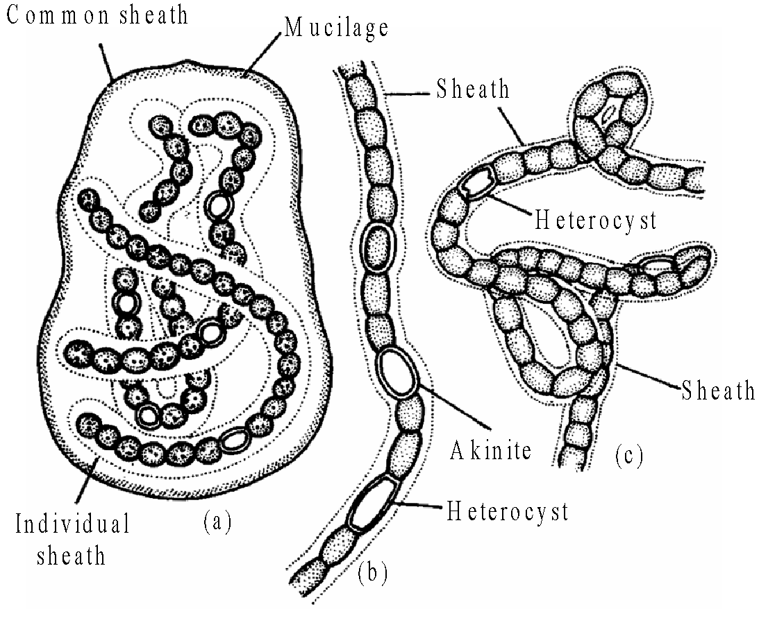
Fig. : Nostoc
- Chemosynthetic autotrophic bacteria oxidize various inorganic substances such as nitrates, nitrites and ammonia and use the released energy for their ATP production. They play a great role in recycling nutrients like nitrogen, phosphorous, iron and sulphur.
- Heterotrophic bacteria are most abundant in nature. The majority are important decomposers. Many of them have a significant impact on human affairs. They are helpful in making curd from milk, production of antibiotics, fixing nitrogen in legume roots, etc. Some are pathogens causing damage to human beings, crops, farm animals and pets. Cholera, typhoid, tetanus, citrus canker are well known diseases caused by different bacteria.
- Bacteria reproduce mainly by fission. Sometimes, under unfavourable conditions, they produce spores. They also reproduce by a sort of sexual reproduction by adopting a primitive type of DNA transfer from one bacterium to the other.
MYCOPLASMA
- Mycoplasmas are organisms that completely lack a cell wall. These are the smallest living cells known and can survive without oxygen. Many Mycoplasma are pathogenic in animals and plants.
- Unit membrane is made up of lipoprotein. The genetic material is a single, linear, double stranded molecule of DNA, without a nuclear envelope.
- Mycoplasma hominis causes pleuropneumonia, inflammation of genitals and endocarditis, etc. Mycoplasma pneumoniae causes PAP (primary atypical pneumonia), haemorrhagic, laryngitis, etc. Mycoplasma fermentatus and M. hominis cause infertility in man, otitis media (inflammation of the middle ear).
- Mycoplasma mycoides causes pneumonia in cattle. Mycoplasma bovigenitalum causes inflammation of genitals in animals. Mycoplasma agalactiae causes agalactia of sheep and goat.
- Common mycoplasmal diseases of plants are : Bunchy top of papaya, witches’ broom of legumes, yellow dwarf of tobacco, stripe disease of sugarcane, little leaf of brinjal, clover phyllody, big bud of tomato etc.
KINGDOM PROTISTA
- All single-celled eukaryotes are placed under Protista, but the boundaries of this kingdom are not well defined.
- Members of protista are primarily aquatic. This kingdom forms a link with the others dealing with plants, animals and fungi. Being eukaryotes, the protistan cell body contains a well defined nucleus and other membrane-bound organelles. Some have flagella or cilia.
- Protists reproduce asexually and sexually by a process involving cell fusion and zygote formation. It may be photosynthetic, holotropic, saprotrophic, parasitic and symbionts. Some have mixotrophic nutrition (holotropic + saprobic). The photosynthetic, floating protists are collectively called phytoplankton. The free-floating, holozoic protozoans are collectively termed zooplankton.
- Unicellular protists have been broadly divided into three major groups :
- Photosynthetic protists : e.g., dinoflagellates, diatoms, euglenoids.
- Consumer protists : e.g., slime moulds or myxomycetes.
- Protozoan protists : e.g., zooflagellata, sarcodina, sporozoa, ciliata.
CHRYSOPHYTES
- This group includes diatoms and golden algae (desmids).
- They are found in fresh water as well as in marine environments. They are microscopic and float passively in water currents (plankton).
- The reserve food material is oil and a polysaccharide-chrysolaminarin (or leucosin).
- In diatoms, the cell walls form two thin overlapping shells, which fit together as in a soap box. The walls are embedded with silica and thus, the walls are indestructible. Thus, diatoms have left behind large amounts of cell wall deposits in their habitat; this accumulation over billions of years is referred to as ‘diatomaceous earth’. Being gritty, this soil is used in polishing, filtration of oils and syrups. Diatoms are the chief ‘producers’ in the oceans.
DINOFLAGELLATES
- These organisms are mostly marine and photosynthetic.
- They appear yellow, green, brown, blue or red depending on the main pigments present in their cells. The cell wall has stiff cellulose plates on the outer surface.
- Most of them have two flagella; one lies longitudinally and the other transversely in a furrow between the wall plates. Very often, red dinoflagellates (Example: Gonyaulax) undergo such rapid multiplication that they make the sea appear red (red tides). Toxins released by such large numbers may even kill other marine animals such as fishes.
- The reserve food material is starch in fresh water forms and oil in marine forms.
- Dinoflagellates reproduce asexually through cell division or by the formation of zoospores and cysts.
- If sexual reproduction occurs, it is isogamous or anisogamous. Two cells conjugate by a conjugation canal where the two amoeboid gametes fuse to form a diploid zygote. Life cycle involves zygotic meiosis (e.g., Ceratium, Gymnodinium etc.) or gametic meiosis (e.g., Noctiluca).
EUGLENOIDS
- Majority of them are freshwater organisms found in stagnant water.
- These protists are devoid of cellulose cell wall. The body is covered by thin and flexible pellicle.
- They have two flagella, a short and a long one. Though they are photosynthetic in the presence of sunlight, when deprived of sunlight they behave like heterotrophs by predating on other smaller organisms. Interestingly, the pigments of euglenoids are identical to those present in higher plants. Example: Euglena.
- The two flagella join with each other at a swelling called paraflagellar body. An orange-red coloured eyespot or stigma is located at the base of flagellum attached to the membrane of the reservoir at the level of paraflagellar body. They contain red pigment astaxanthin. Both paraflagellar body and eye spot act as photoreceptors and direct the organism towards the optimum light.
- Sexual reproduction has not yet been definitely proved. Under favourable conditions, euglenoids multiply by longitudinal binary fission.
- Euglena is a connecting link between animals and plants.
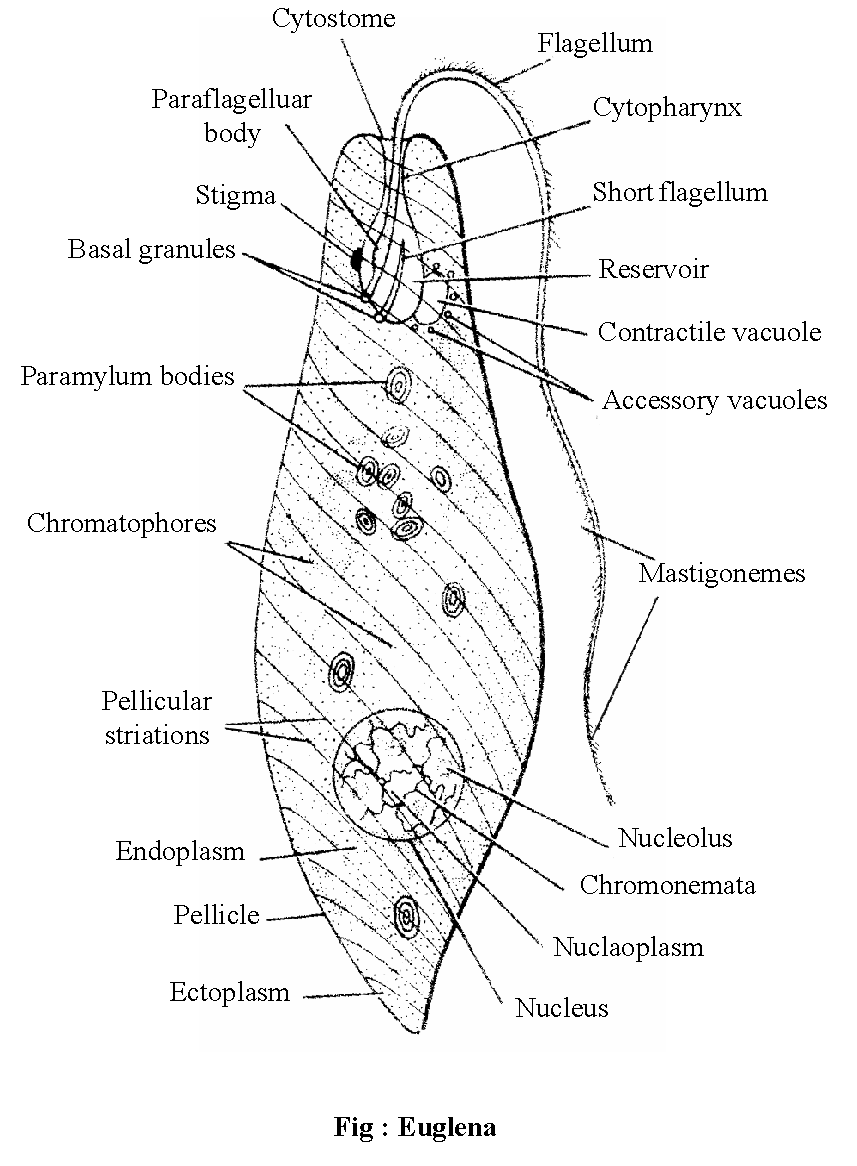
Fig. : Euglena
SLIME MOULDS
- Slime moulds are saprophytic protists. The body moves along decaying twigs and leaves engulfing organic material.
- Under suitable conditions, they form an aggregation called Plasmodium which may grow and spread over several feet. During unfavourable conditions, the Plasmodium differentiates and forms fruiting bodies bearing spores at their tips. The spores possess true walls. They are extremely resistant and survive for many years, even under adverse conditions. The spores are dispersed by air currents.
- Slime moulds are of two types : acellular and cellular
ACELLULAR (PLASMODIAL) SLIME MOULDS
- Acellular slime moulds commonly grow as slimy masses on damp places rich in dead and decaying organic matter.
- The somatic phase is diploid and consists of a free living organic matter with multinucleated protoplasm called plasmodium.
- The Plasmodium slowly streams or glides over decaying organic matter putting out blunt finger like pseudopodia showing amoeboid movement.
- They also absorb dissolved organic substances from the substratum showing saprotrophic nutrition.
- Under unfavourable conditions, the plasmodium contracts and gets surrounded by thick horny wall. It is called sclerotium.
- Each plasmodium reproduces asexually by the formation of several, small, sessile or stalked, brightly coloured sporangia.
- The multinucleated protoplasm of sporangium is cleaved to produce a large number of small uninucleate spores.
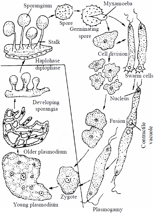
Fig. : Life cycle of Acellular Slime mould (e.g., Physarum)
CELLULAR SLIME MOULDS
- The cellular slime moulds occurs in the form of haploid uninucleated, naked (without cell wall) cell covered by plasma membrane. These cells are called myxamoebae or swarm cells.
- The myxamoebae move freely with the help of amoeboid movement and phagotrophic or holozoic nutrition.
- They grow and divide to form a large population of individuals.
- Under unfavourable conditions, a myxamoebae secrete a rigid cellulose wall to form the microcyst. Microcyst formation is a means of perennation.
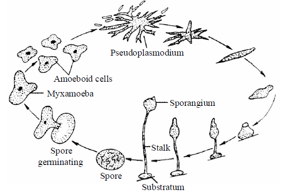
Fig. : Life cycle of cellular slime mould
PROTOZOANS
- Protozoa is the 3rd largest phylum. It is one celled body performed all the biological activities like multicellular animals. So they are termed as "acellular" organism, proposed by Dobell.
- Protozoans were first studied by Leeuwenhoek and the name protozoa was coined by Goldfuss.
- Study of protozoans is known as Protozoology.
- Protozoa are world wide, cosmopolitan mostly microscopic, aquatic, terrestrial , free living (Amoeba) or parasitic (Plasmodium), solitary or colonial (Proterospongia).
- Body level of organisation of protozoans is protoplasmic level. It consists of uninucleate or multinucleate protoplasm mostly naked or some have body bounded by delicate membrane or a firm pellicle; test, lorica or shell. In few groups of protozoa CaCO3 & silica shell's exoskeleton is found. E.g. Radiolarian group & foraminifera group.
- Number of nuclei vary from one to many. Few show nuclear dimorphism, e.g. Paramecium. Body performs all necessary biological activity so in them subcellular-physiological division of labour is found.
- Locomotion is by means of
- Finger-like Pseudopodia, e.g. Amoeba.
- Whip like Flagella, e.g. Euglena.
- Hairy cilia, e.g. Paramecium
- By contraction
- No motion
- Nutrition of protozoans are mainly holozoic (Amoeba), mixotrophic (Euglena), parasitic, saprozoic (Plasmodium) and digestion is intracellular which take place in food vacuole.
- Respiration and excretion take place by exchange of gases through body surface. Some excretion may occur through contractile vacuole.
Nitrogenous waste is ammonia. Some freshwater protozoans get rid of excess water through 'contractile vacuole known as osmoregulation. Amoeba has one and Paramecium has two vacuoles.
- Reproduction takes place by asexual & sexual method
- Asexual reproduction by Binary fission (Amoeba), Transverse fission (Paramecium), Longitudinal fission (Trypansoma, Euglena), Multiple fission (Plasmodium), Budding (Amoeba)
- Sexual reproduction by syngamy (Plasmodium) and conjugation (Paramecium). Some also form cysts which help in unfavourable condition for reproduction of an organism. They do not have natural death because in unicellular animals there is no division of somatoplasm & germplasm so these are considered as immortal.
- Protozoa are divided into four major groups on the basis of locomotory organelles.
- Amoeboid protozoans
- Flagellated protozoans
- Ciliated protozoans
- Sporozoans
AMOEBOID PROTOZOANS
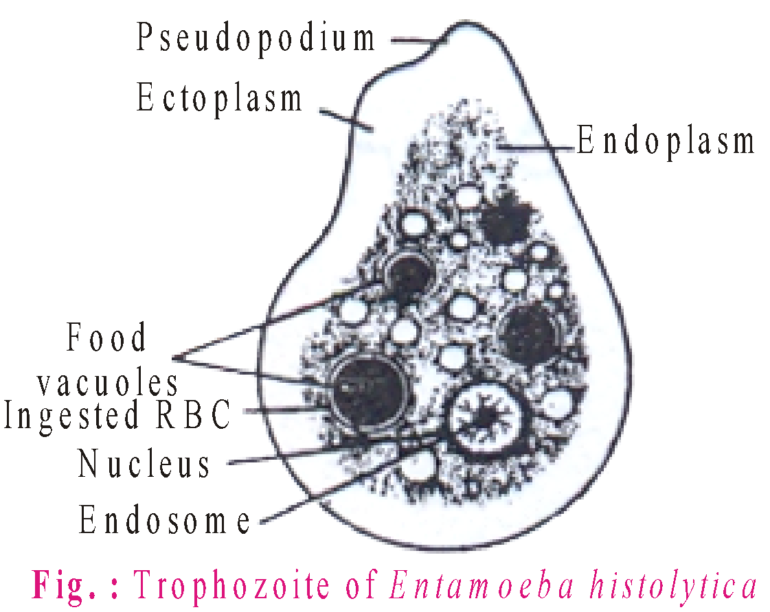
- These organisms live in freshwater, sea water or moist soil. They move and capture their prey by putting out pseudopodia (false feet) as in Amoeba. Marine forms have silica shells on their surface. Some of them such as Entamoeba are parasites.
- Amoeba belongs to the class sarcodina or rhizopoda of the phylum protozoa.
- The most common species is Amoeba proteus. Proteus is the name of the mythical sea god who could change shape.
- Body is covered by plasmalemma. It is a trilaminar and selectively permeable membrane. Plasmalemma is excretory, ammonia diffuses out through it. It is also respiratory as diffusion of oxygen and carbon dioxide takes place through it.
- Pseudopodia are found in Amoeba and leucocyte of higher animals.
- Locomotion of Amoeba is known as amoeboid movement.
- Digestion in Amoeba is intracellular. Amoeba secretes digestive enzymes for hydrolysing starch, protein, fat etc.
- Food vacuole of Amoeba is analogous to the alimentary canal of an animal or gastrovascular cavity of Hydra. The contents of food vacuole in Amoeba first becomes acidic and alkaline.
- Amoeba responds to environmental conditions. Response to the stimuli is called taxis. Different taxis are thermotaxis (temperature), phototaxis (light), thigmotaxis (touch), chemotaxis (chemicals), galvanotaxis (electric current), geotaxis (gravity) and rheotaxis (water current).
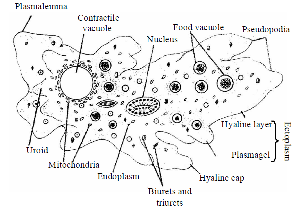
Fig. : Amoeba
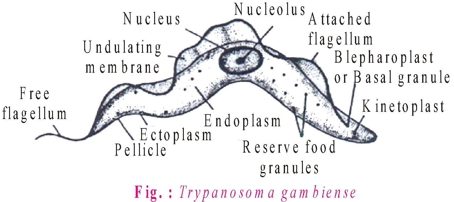
FLAGELLATED PROTOZOANS
- The members of this group are either free-living or parasitic. They have flagella.
- Trypanosoma gambiense is the parasitic zooflagellate which causes one of the deadliest ailments in human beings called African sleeping sickness or Trypanosomiasis. It was discovered by Frode in 1901.
- Trypanosoma is usually found in the blood of vertebrates, finally invading cerebrospinal fluid.
- Trypanosoma reproduces asexually by longitudinal binary fission. It does not form cysts.
- Trypanosoma is an endoparasite, blood parasite, extracellular parasite.
- Trypanosoma is digenetic, that is, it completes its life cycle in two hosts. The primary or principal or definite host is man and the intermediate or secondary host or vector is the insect, tse-tse fly or bug.
CILIATED PROTOZOANS
- These are aquatic, actively moving organisms because of the presence of thousands of cilia. Example: Paramecium.
- Paramecium is commonly called as ‘Slipper animalcule’. Body is distinguished into an oral or ventral surface and an aboral or dorsal surface.
- Body is covered with a thin, firm, flexible membrane called pellicle. Entire body surface is covered by numerous cilia, the locomotory organelles.
- Digestion in Paramecium is intracellular. Food vacuole constantly moves along a definite course (cyclosis) within streaming endoplasm. Food vacuole is digested in the cell body in acidic to alkaline media. Egestion of undigested food takes place through cytopyge or cytoproct, a temporary formed anus.
- Paramecium reproduces by transverse binary fission and nuclear reorganisation. Binary fission occurs during favourable conditions. In this process, macronucleus divides amitotically and micronucleus mitotically.
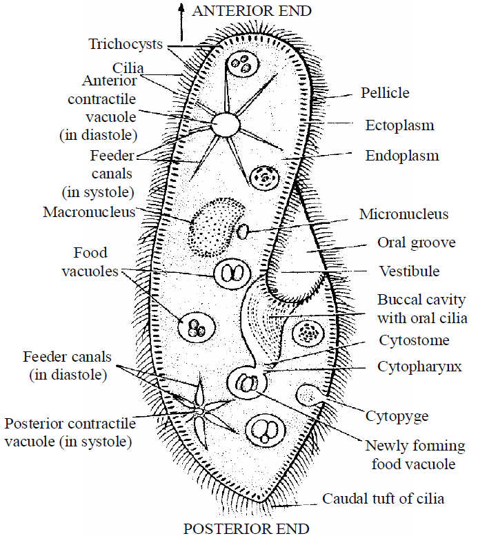
Fig. : Paramecium
SPOROZOANS
- These are spore forming parasitic protists which lack locomotory structure and contractile vacuoles. The body is covered by a pellicle or cuticle.
The most notorious is Plasmodium (malarial parasite) which causes malaria having a staggering effect on human population.
- Laveran (1880) discovered that malaria is caused by a protozoan parasite, Plasmodium vivax. Sir Ronald Ross (1896) was the first to observe oocytes of Plasmodium in female Anopheles.
- In the life cycle of Plasmodium, two important phases are present.
- Endogenous or Asexual phase : passes in man.
- Exogenous or Sexual phase : passes in female Anopheles mosquito.
KINGDOM FUNGI
- The fungi are a group of eukaryotic microorganisms that lack chlorophyll, are unable to synthesize their own food and are therefore heterotrophic.
- The branch of science that deals with the study of fungi is called Mycology.
- Fungi possess all eukaryotic organelles and reserve food particles (glycogen, lipids etc.)
- With the exception of yeasts which are unicellular, fungi are filamentous. Their bodies consist of long, slender thread-like structures called hyphae. The network of hyphae is known as mycelium. Some hyphae are continuous tubes filled with multinucleated cytoplasm – these are called coenocytic hyphae. Others have septate or cross walls in their hyphae.
- The cell walls of fungi are composed of chitin and cellulose. While, chitin is a polymer of N-acetyl glucosamine, cellulose is a polymer of D-glucose.
- Those fungi that depend on living plants and animals are called parasites. They can also live as symbionts – in association with algae as lichens and with roots of higher plants as mycorrhiza.
- Fungi possess true nucleus having definite nuclear envelope. The nuclear envelope persists during nuclear division.
- The fungi reproduce by all the three methods - vegetative, asexual and sexual.
- Reproduction in fungi can take place by vegetative means – fragmentation, fission and budding.
- Asexual reproduction is by spores called conidia or sporangiospores or zoospores, and sexual reproduction is by oospores, ascospores and basidiospores. The various spores are produced in distinct structures called fruiting bodies.
- The sexual cycle involves the following three steps:
- Fusion of protoplasms between two motile or non-motile gametes called plasmogamy.
- Fusion of two nuclei called karyogamy.
- Meiosis in the zygote resulting in haploid spores.
- When a fungus reproduces sexually, two haploid hyphae of compatible mating types come together and fuse. In some fungi, the fusion of two haploid cells immediately results in diploid cells (2n). However, in other fungi (ascomycetes and basidiomycetes), an intervening dikaryotic stage (n + n i.e. two nuclei per cell) occurs; such a condition is called a dikaryon and the phase is called dikaryophase of fungus. Later, the parental nuclei fuse and the cells become diploid. The fungi form fruiting bodies in which reduction division occurs, leading to the formation of haploid spores.
- The classification of fungi based on the characteristics of the life cycle involved like nature of somatic phase, kinds of asexual spores, kinds of sporangia, nature of the life cycle and presence or absence of perfect or sexual stage.
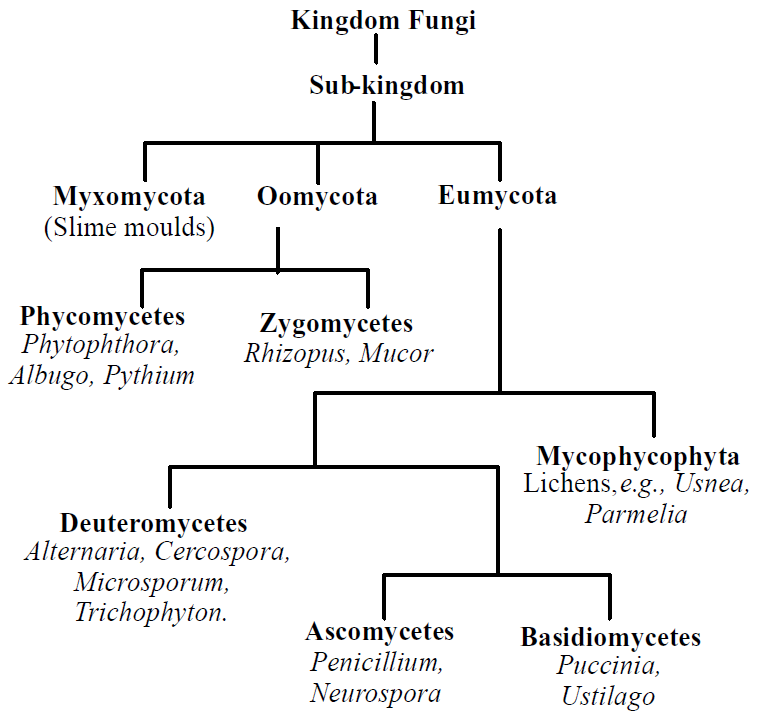
Flow chart 2.7 : Classification of Fungi
PHYCOMYCETES
- Phycomycetes are algae like fungi.
- Members of phycomycetes are found in aquatic habitats and on decaying wood in moist and damp places or as obligate parasites on plants. The mycelium is aseptate and coenocytic.
- Two types of flagella are present in phycomycetes, these are whiplash and tinsel type.
- Asexual reproduction takes place by zoospores (motile) or by aplanospores (non-motile). These spores are endogeneously produced in sporangium. Zygospores are formed by fusion of two gametes. These gametes are similar in morphology (isogamous) or dissimilar (anisogamous or oogamous). Examples : Mucor , Rhizopus and Albugo (the parasitic fungi on mustard).
- Rhizopus / Mucor - They are cosmopolitan and saprophytic fungus, living on dead organic matter. Rhizopus stolonifer occur very frequently on moist bread, hence commonly called black bread mold.
- Mucor is called dung mold. Both are called black mold or pin mold because of black coloured pin head like sporangia. Besides, it appears in the form of white cottony growth on moist fresh organic matter, jams, jellies, cheese, pickles, etc.
- These reproduce by vegetative, asexual and sexual methods.
- Vegetative reproduction takes place by fragmentation. If stolon breaks accidentally into small segments, each part grows into a new mycelium.
- Asexual reproduction occurs by three types of non-motile mitospores, that is sporangiospores, chlamydospores and oidia.
- Sexual reproduction takes place by conjugation between two multinucleate but single celled gametangia. The gametes are isogamous and non-motile.
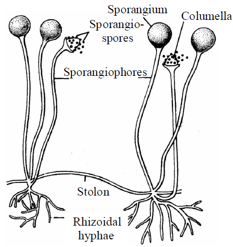
Fig. : Rhizopus
ECONOMIC IMPORTANCE
- Spoilage of food : Exposed bread and other food particles are spoiled by Rhizopus and Mucor species.
- Soft rot : Rhizopus species attack sweet potato, apple and strawberry producing soft rot or leak disease. Germinating maize grains are also attacked.
- Mucormycosis : Mucor pusillus and M. ramosissimus may attack internal human organs, including lungs, alimentary canal and nervous system.
- Fermented foods : Tempeh (a solid food from soyabean) and sufu (Chinese cheese) are prepared with the help of Rhizopus and Mucor respectively.
- Chemicals : Citric acid prepared by Mucor from molasses, fumaric acid and cortisone by Rhizopus stolonifer, lactic acid by R. stolonifer and R.nodosus and alcohol by R. oryzae and M. javanicus.
- Antibiotic : Ramysin is produced by Mucor ramannianus.
- Wastewater treatment : Growth of Mucor arrhizus removes heavy metal contamination of water.
ALBUGO
- Albugo is an obligate parasite and grows in the intercellular spaces of host tissues.
- It is parasitic mainly on the members of families cruciferae, compositae, amaranthaceae and convolvulaceae. The disease caused by this fungus is known as white rust or white blisters.
- The most common and well known species is Albugo candida which attacks the members of the mustard family (Cruciferae). It is commonly found on Capsella bursa pastoris (Shepherd’s purse) and occasionally on radish, mustard, cabbage, cauliflower, etc. The reserve food is oil and glycogen.
ASCOMYCETES
- These are unicellular as well as multicellular fungi. In the latter, mycelium is septate.
- The asexual spores formed in chains are called conidia. These detach from the parent and form new mycelia.
- The asexual spores are conidia produced exogenously on the special mycelium called conidiophores. Conidia on germination produce mycelium. Sexual spores are called ascospores which are produced endogenously in sac like asci (singular ascus). These asci are arranged in different types of fruiting bodies called ascocarps.
- Some examples are Aspergillus, Claviceps and Neurospora. Neurospora is used extensively in biochemical and genetic work. It is known as Drosophila of plant kingdom. Many members like morels and buffles are edible and are considered delicacies.
- Yeast was first described by Antony Von Leeuwenhoek in 1680. Yeast are non-mycelial or unicellular, small and either spherical or oval in shape.
- Under favourable conditions, yeast grow rapidly and form false mycelium or pseudomycelium. Individual cells are colourless but the colonies may appear white, red, brown, creamy or yellow.
- The single cell of yeast is about 10 μm in diameter. It is enclosed in a delicate membrane which is not made up of fungal cellulose but is a mixture of two polysaccharides known as mannan and glycogen.
- Yeast reproduces by vegetative or asexual and sexual methods.
- Yeast reproduce vegetatively either by budding or by fission.
- Sexual reproduction in yeasts takes place during unfavourable conditions, particularly when there is less amount of food.
The sex organs are not formed in yeasts and the sexual fusion occurs between the two haploid vegetative cells or two ascospores which behave as gametes. The two fusing gametes are haploid and may be isogamous or anisogamous. Such kind of sexual reproduction is called gametic copulation. It is the best example of hologamy i.e., the entire vegetative thallus is transformed into reproductive body. The sexual fusion leads to the formation of diploid zygote. The zygote behaves as an ascus and forms 4 – 8 haploid ascospores. These liberate and function as vegetative cells.
ECONOMIC IMPORTANCE
- Useful activities
- Baking industry : Yeast are used in the manufacture of bread. Kneaded flour is mixed with yeast and allowed to ferment. Yeast convert starch into sugars and sugar into CO2 and alcohol with the help of enzyme zymase. CO2 is released when effervescence takes place due to which bread becomes spongy and gets swollen and is of light weight.
- Brewing industry : Brewer’s yeast or Beer yeast is Saccharomyces cerevisiae and wine yeast is Saccharomyces ellipsoideus. These perform alcoholic fermentation.
- Food yeast : Yeast from brewing industry is harvested and used as food yeast. It is rich in protein and vitamin-B (Riboflavin).
- Harmful activities
- Fermentation of fruits and fruit juices by yeast cells makes their taste unpleasant.
- Parasitic species of yeast like Nematospora causes diseases in tomato, cotton and bean.
- Parasitic yeast cause diseases in human beings (e.g., cryptococcosis, blastomycosis and torulosis)
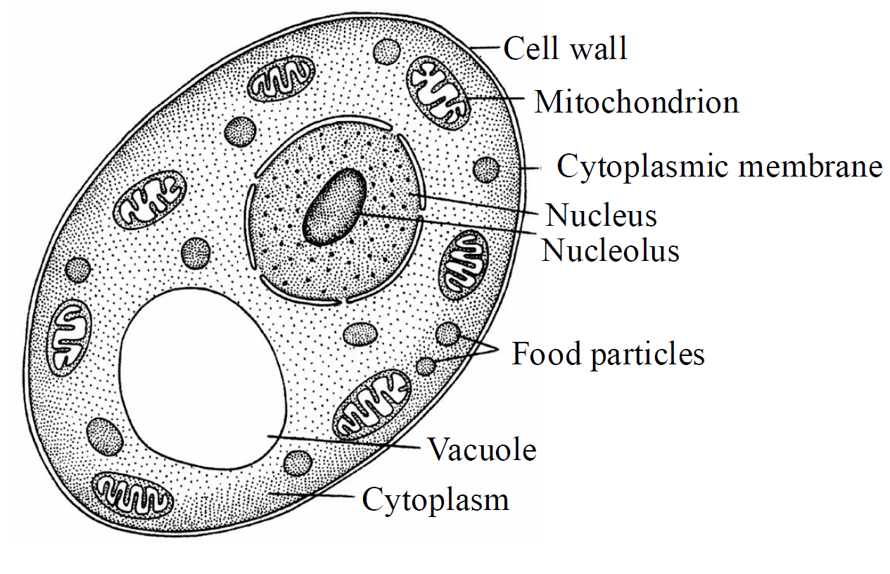
Fig. : Yeast Cell
BASIDIOMYCETES
- Basidiomycetes are commonly known as club fungi.
- It resembles the ascomycetes in having a septate mycelium and production of non-motile spores.
- Commonly known forms of basidiomycetes are mushrooms, bracket fungi or puffballs. They grow in soil, on logs and tree stumps and in living plant bodies as parasites, e.g., rusts and smuts. The mycelium is branched and septate.
- The asexual spores are generally not found, but vegetative reproduction by fragmentation is common. The sex organs are absent, but plasmogamy is brought about by fusion of two vegetative or somatic cells of different strains or genotypes. The resultant structure is dikaryotic which ultimately gives rise to basidium. Karyogamy and meiosis takes place in the basidium producing four basidiospores. The basidiospores are exogenously produced on the basidium. The basidia are arranged in fruiting bodies called basidiocarps.
- The dikaryotic cells divide by clamp connections. A lateral pouch or clamp like outgrowth arises which projects downward like a hook. The two nuclei now undergo conjugate division in such a way that-one spindle lies parallel to the long axis of the cell and the other somewhat obliquely. As a result, one daughter nucleus enters into the clamp.
- Some common members are Agaricus (mushroom), Ustilago (smut) and Puccinia (rust fungus).
DEUTEROMYCETES
- It is commonly known as imperfect fungi because only the asexual or vegetative phases of these fungi are known.
- The deuteromycetes reproduce only by asexual spores known as conidia. The mycelium is septate and branched. Some members are saprophytes or parasites while a large number of them are decomposers of litter and help in mineral cycling.
- Examples : Alternaria, Colletotrichum, Trichoderma, Fusarium, Helminthosporium oryzae, Cercospora personata.
SOME COMMON NAMES OF FUNGI
- Myxomycetes - Slime fungi or slime or mycocetozoa
- Eumycetes - True fungi
- Phycomycetes - Algal fungi or aquatic fungi
- Ascomycetes - Sac fungi
- Basidiomycetes - Club fungi (e.g. Saprolegnia)
- Allomyces - Water molds or aquatic fungi (e.g. Blastocladiella)
- Pythium - Damping off fungus
- Mucor - Black or Bread mold
- Mucor mucedo - Dung mold
- M. Stolonifer - Bread mold
- Penicillium - Green or blue mold
- Pezzia - Cup fungi
- Polyporus - Pore fungi
- Morchella - Morel or sponge mushroom
- Agaricus campestris - Common edible mushroom
- Poisonous mushroom - Toadstool
- Coprinus comatus - Inky cap mushroom
- Synchytrium - Chytrid
- Aspergillus - Conidial fungus
- Neurospora - Red bread mold or Bakery mold or Genetical fungus or Drosophila of plant kingdom
- Puccina - Rust fungus
- Ustilago - Smut fungus
- Lycoperdon - Puffball or Earth star
- Cyathus, Nidula - Bird’s nest fungi
- Nidularia - Fairy purse
- Pernospora - Downy mildew
- Aspergillus niger - Black mold or weed of laboratory
- Saccharomcyes crevisiae - Yeast of commerce
- Marasimus oreaders - Fairy rings fungi
- Yeast - SCP, sprouting fungi, sugar fungi
- Mucor stolonifer - Bread mold
- Claviceps - Ergot fungi
- Marchella - Edible sac fungi
- Auricularia - Jelly fungi
- Dictyophora - Stinkhorn fungi duplicate
- Fomes - Shelf fungi
- Clavicornona - Coral fungi
- Armillariella mellea - Honey mushroom
- Psilocybe geastrum - Hallucinogenic fungi
- Astraeus - Earth stars
- Labryrinthula - Net slime molds
- Rhizopus - Conjugation fungi
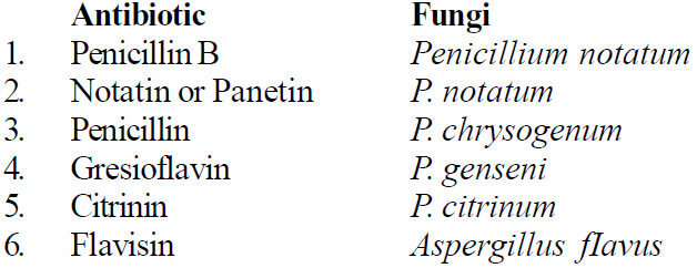
SOME IMPORTANT ANTIBIOTICS
KINGDOM PLANTAE
- Kingdom plantae includes all eukaryotic chlorophyll-containing organisms commonly called plants.
- Cells are surrounded by cell wall and contain cellulose.
- Reserve food is starch in green algae and embryophytes, floridean starch in red algae and laminarian in brown algae.
- Growth occurs due to the presence of definite growing points or cells. In higher forms, growing areas are called meristems.
- A multicellular embryo is formed during development from the zygote. Life cycle consists of alternating haploid gametophyte and diploid sporophyte generation. This phenomenon is called alternation of generations.
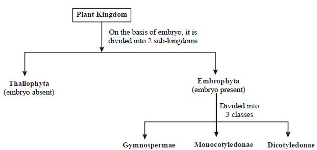
- August Wilhelm Eichler (1883), a Vinnese botanist, divided plant kingdom into two sub-kingdoms mainly on the basis of presence or absence of seeds.
- Cryptogamae are lower plants in which sex organs are hidden and seeds and flowers are absent. It includes thallophytes, bryophytes, pteridophytes.
- Phanerogamae are higher plants in which sex organs are evident; seeds present. It includes gymnosperms and angiosperms.
- In modern system of classification like Whittaker (1969), fungi, lichens and bacteria are excluded from this group and are placed in separate kingdoms.
KINGDOM ANIMALIA
- This kingdom is characterized by heterotrophic eukaryotic organisms that are multicellular and their cells lack cell walls.
- Animals have heterotrophic mode of nutrition.
- They require oxygen for aerobic respiration.
- Animals are able to make rapid responses to external stimuli as a result of the activity of nerve cells, muscle or contractile tissue or both.
- Animal life cycle includes stages of embryonic development. Mitotic cell divisions (cleavage) transform the animal zygote into a multicellular embryo.
- Anaemia are animals without red blood, e.g., sponges, cnidaria, mollusca, arthropoda, echinodermata, etc.
- Enaima are animals with red blood, e.g., vertebrates.
- Vivipara are those animals which give birth to young ones, e.g., man, dogs, cows, etc.
- Ovipara are those animals which lay eggs, e.g., frogs, toads, lizards, snakes, birds, etc.
- Anamniotes are vertebrates without embryonic membranes e.g., fishes, amphibians.
- Amniotes are vertebrates with embryonic membranes (chorion, amnion, allantois, yolk sac) e.g., reptiles, birds, mammals.
- Acraniata or protochordata are chordates without cranium (brain box). It includes urochordata and cephalochordata.
- Chordates are animals with notochord, dorsal tubular nerve cord, paired pharyngeal gill slits. All urochordates, cephalochordates and vertebrates are called chordates.
- Craniata or vertebrates are chordates with cranium. It includes cyclostomes, pisces, amphibians, reptiles, birds and mammals.
- Non-chordates are animals without a notochord (a rod like elastic structure which supports the body). Phylum porifera to phylum hemichordata are called non-chordates.
- Invertebrates are animals without vertebral column (backbone). All the non-chordates, urochordates and cephalochordates are collectively called invertebrates.
VIRUSES, VIROIDS AND LICHENS
- The term 'virus' has been derived from Latin, which means poison or venom or viscous fluid. They remain inactive outside a living host but become active inside the host and multiply in it.
- D.J. Ivanowsky (1892) recognised certain microbes as causal organism of the mosaic disease of tobacco. These were found to be smaller than bacteria because they passed through bacteria-proof filters.
- M.W. Beijerinek (1898) demonstrated that the extract of the infected plants of tobacco could cause infection in healthy plants and called the fluid as Contagium vivum fluidum (infectious living fluid).
- W.M. Stanley (1935) showed that viruses could be crystallised and crystals consist largely of proteins. They are inert outside their specific host cell.
- In addition to proteins, viruses also contain genetic material, that could be either RNA or DNA.
- A virus is a nucleoprotein and the genetic material is infectious. In general, viruses that infect plants have single stranded RNA and viruses that infect animals have either single or double stranded RNA or double stranded DNA.
- Bacterial viruses or bacteriophages (viruses that infect bacteria) are usually double stranded DNA viruses and contain lysozyme enzyme.
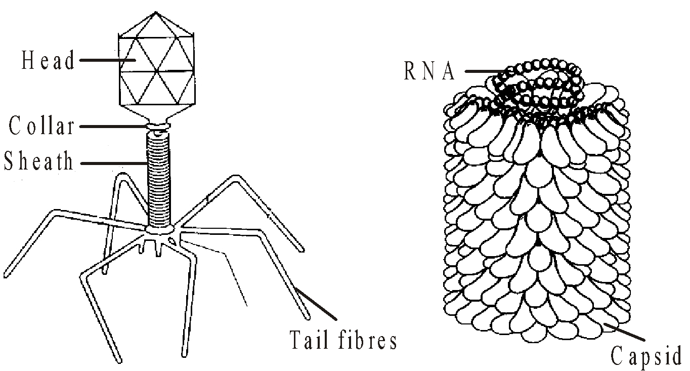
Fig. : (a) Bacteriophage; (b) Tobacco Mosaic Virus (TMV)
- The protein coat called a capsid made of small subunits called capsomeres, protects the nucleic acid. These capsomeres arranged in helical or polyhedral geometric forms possess antigenic properties.
- Viruses cause diseases like mumps, smallpox, herpes and influenza. AIDS in humans is also caused by a virus. In plants, the symptoms can be mosaic formation, leaf rolling and curling, yellowing and vein clearing, dwarfing and stunted growth.
- In 1971, T.O. Diener discovered very simple smallest infectious agents called Viroids. They consist of RNA and capsid is lacking. The RNA of the viroid was of low molecular weight. They cause persistent infections.
- A lichen is structurally organised entity consisting of the permanent association of a fungus and an alga. The algal component is known as phycobiont and fungal component as mycobiont, which are autotrophic and heterotrophic, respectively.
- Algae prepare food for fungi and fungi provide shelter and absorb mineral nutrients and water for its partner.
- Lichens reproduce both by asexual and sexual methods.
- Lichens are very sensitive to SO2 and grow only in SO2 free atmosphere.
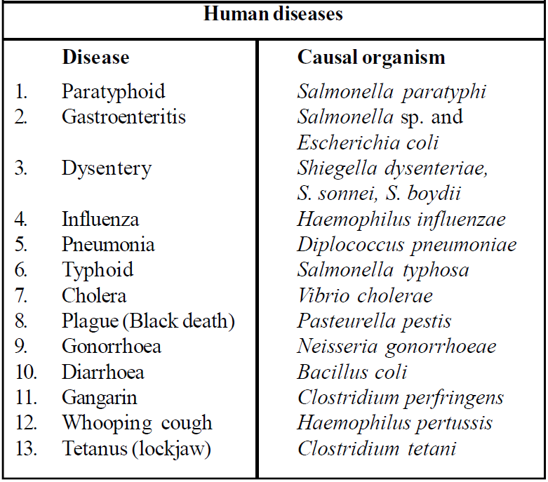
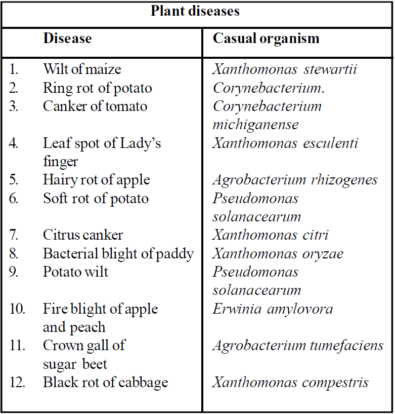
Table : Diseases and their causal organisms
Study Notes for NEET/AIIMS/JIPMER


